
Cancer cachexia
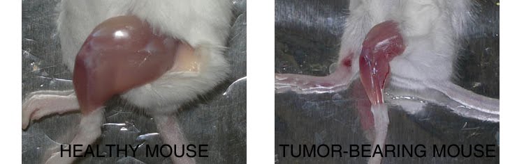
Compared to a control mouse (left) a tumor-bearing mouse (right) displays a dramatic muscle wasting. This loss of muscle mass is called cancer cachexia.
Exogenous gene expression in regenerating muscle
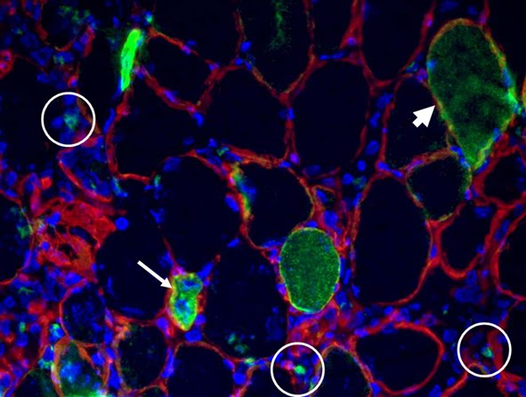
Depicted here is the over-expression of Green Fluorescent Protein (GFP, green; click on the image to access Tsien's Lab) in interstitial cells (circled), nascent myofibers (arrow) and adult fibers (arrowhead), in a regenerating Tibialis Anterior following focal injury. Laminin staining (red) highlights the basement membrane surrounding the skeletal muscle tissue, while nuclei are stained in blue. We do gene delivery by electroporation to study the regulation of muscle regeneration.
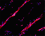
Cultures of myotubes on a conductive surface in a parallel orientation.
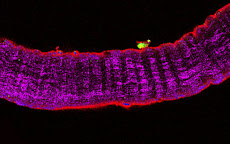
C2C12 cells cultured on gold, by mean of adhesion to 100 nm-wide stripes coated with anti Stem Cell antigen1 (Sca1) Ab. Nuclei (blue) and actin cytoskeleton (red) staining highlights the selective cells adhesion on the Ab-coated stripes and the formation of parallel multinucleated syncytia (myotubes).
3/21/2023
Sarcopenic obesity: where to go
2023 - Comment article published in Clinical Nutrition, suggesting the need for future research on sarcopenic obesity.
Sarcopenic obesity research perspectives outlined by the Sarcopenic Obesity Global Leadership Initiative (SOGLI) – proceedings from the SOGLI Consortium meeting in Rome November 2022
I participated to the meeting as an expert on sarcopenia.
Below please find the link to access the fulltext
Elsevier Share Link: https://authors.elsevier.com/c/1gnOuxUE3j7FZ
Sarcopenic obesity has health consequences ultimately leading to functional decline and increased risks of cardiometabolic diseases and mortality. The European Working Group on Sarcopenia in Older People (EWGSOP) developed a practical clinical definition and consensus diagnostic criteria for age-related sarcopenia: the consensus paper on definition and diagnosis can be found here (Cruz-Jentoft et al. Age Ageing. 2010 Jul;39(4):412-23. doi: 10.1093/ageing/afq034. PMID: 20392703)
 The SOGLI group has now identified the most relevant avenues or future research on sarcopenic obesity, in order to fill the gap on the most relevant aspects of this condition, which is not just the sum of age-related sarcopenia and obesity. The read more click here
4/20/2022
In this world we are a family
A video from our students at Sapienza in a diffcult moment for our world ----- International Medical School - 2nd y students: "In this world..." (2022)
6/04/2021
All classes of my course of Introduction to Research and Scientific Method
Below, you will find my classes (for the full slides, follow the link in the title of each class, below) of my course: Introduction to Research and Scientific Method, for the 1st y college students at Sorbonne University. Below is the planning. 20 hours, 3 credits. The course is mostly in French.
1 PRÉSENTATION: DEMARCHE SCIENTIFIQUE
Introduction de la thématique: comment réussir un travail universitaire
Le muscle squelettique et ses disciplines
La démarche scientifique" Se documenter sur le sujet 'expérimentation animale' de manière à pouvoir en discuter (travail à la maison; individuel)
2 RECHERCHE BIBLIOGRAPHIQUE
Présentation de la méthodologie
Présentation des resources @ l’UPMC
TD" TD bibliographique (sur place et à la maison; individuel)
3 MODELES EXPERIMENTAUX
Introduction de la thématique
modèles expérimentaux
niveaux d’observation et organismes modèles
demande d'autorisation à l’experim. anim.
débat: expérimentation animale" "Choisir un article
scientifique (entre ces proposés sur Sakai en S3) et rédiger
une fiche de lecture (à
la maison; en trinôme):
indiquer dans la fiche mots les clés thématiques, le(s) modèle(s), ainsi que
les méthodes.Etudier les annexes S3 fichier Lentz + fichier Neveu"
4 MÉTHODE EXPÉRIMENTALE
Reprise «Modèle animal»: la souris
Introduction de la thématique: l’analyse des données et l'interprétation
Observation, contrôles et artefacts
le cahier du laboratoire
un exemple réel: activité physique de la souris
Abstract: résumé scientifique" Se préparer à la conference (S5)
5 CONFERENCE
Se préparer sur le sujet de la conference
Suivre la conference, sachant qu'il faudra :
en faire un résumé verison Grand Publique (GP)
comparer l’exposition pour un conference avec celle dans un article de données
prendre des notes sur l’exposé oral et évaluer l’orateur en 5 lignes sur la base de critères de Public speaking (voir S9)" Rédaction d’un Fiche critique de la conférence en Français (une page et demi max), y compris résume en 5 lignes du contenu à l'intention du GP + 5 ligne d’évaluation de l’orateur (à la maison, à envoyer par email; individuel)
6 SAVOIR LIRE: ARTICLES SCIENTIFIQUES
Correction du travail à la maison S2, S3
Introduction de la thématique
L’article des données
Reproductibilité, validation, rétractation
présentation d’un vraie article à évaluer
TD: Real peer review of a submitted article (correction sur place; en groupe)
Cahier des charges, formation de groupes pour la rédaction d’un dossier scientifique" "Recherche d’un sujet
de dossier scientifique: bibliographie et
rédaction du dossier
(à la maison ; en groupe)
7 DOSSIER SCIENTIFIQUE
Correction du travail (S5) à la maison
Organization, discussion du contenu
Comment rédiger un dossier scientifique
Plagiat
Reproductibilité, société, le cas DBM°
Cahier des charges, réunion de groupes pour la rédaction d’un dossier scientifique
° the Di Bella Method " Rédaction du dossier (à la maison ; en groupe)
8 SAVOIR ECRIRE: PROJETS ET RESUMES
Le résumé scientifique: pour un congres, pour une demande de financement
Rédaction de projets: hints and tips
TP: Travail en binome sur le résumé scientifique (abstract) pour un congres, à partir du «cahier du labo» réel de la S4 (en classe; en binôme)
réunion groupes dossier scientifique
" "Résumé scientifique (abstract) à partir du «cahier du labo» (en classe; en binôme)
Rédaction final du dossier selon le format imposé diffusé en cours et sur Sakai (à la maison ; en groupe)
9 SUPPORTS GRAPHIQUES
Correction du travail (S8) à la maison
Introduction de la thématique
Schema vs dessin
Visual Abstracts
Comment faire un bon diaporama
préparation S7 Public Speaking : préparer un discours de 1 min exacte (en Anglais) pour expliquer une idée ou un système (à la maison; individuel)
10 invited speaker: Dr. A. Mazzotti (interpreter & actress)" "SAVOIR PARLER: PUBLIC SPEAKING + COMMENT FAIRE UNA BONNE PRESENTATION (DIAPORAMA)
Introduction de la thématique: l’exposé oral
public speaking : l'oral qui intègre et accompagne le diapos, langage du corps, communication publique
"Réalisation d'un poster en diaporama pour illustrer le dossier ou un article spécifique du dossier;
Préparation de l’oral (à la maison ; en groupe)"
11 "SOUTENANCE DES POSTERS*
Évaluation de l’enseignement
Foire à questions (5 min Qs sur les sujets des présentations)
Restitution des dossiers corrigés" Présentation orale du poster: durée max de l’exposé orale 15 min (en groupe/individuel*)
* poster version globale et divisé en diapo * approx 3 min chacun
4/12/2019
LECTURES, CLASSES ETC: METABOLISM AND CANCER STRIATED MUSCLE RESPONSES ASSOCIATED TO CANCER CACHEXIA
3/12/2017
SEE YOU IN PADUA


2/05/2017
ARTICLES: PubMed Central® or PubMed centrally located?

Smile! Life is beautiful after all
 Dedicated to ILoveHistology, the best website to have fun with histology.

6/03/2016
ARTICLES: Sweating Hard or Swallowing Pills?

ARTICLES: Eating to live or living to eat?


Figure legend. Flaxseed-enriched diet preserves dystrophic skeletal muscle morphology. For all in vivo observations, dystrophic hamsters were fed with a flaxseed- enriched diet (FS) from weaning to the age of 150 days (Dystr/FS) and compared with dystrophic (Dystr/P) and healthy hamsters (Healthy) fed with standard pellet (P). (A) Representative images of H&E-stained skeletal muscle sections. Scale bar: 50 μm. (B) Percentage of myofibers with internalized nuclei from H&E-stained skeletal muscle sections. *P<0.05 vs. Healthy; § P<0.05 vs. Dystr/P; n=5. (C) Component Score Coefficient Matrix. The coefficients by which variables are multiplied to obtain factor scores are shown. The variables are represented by the morphometric parameters derived from light microscope images of skeletal muscle (six sections from each of 5 animals/group). The values highlighted (red boxes) indicate the variables most closely associated with Principal Components 1 and 2. (D) Principal Component Analysis (PCA). Three series of data from of Healthy, Dystr/FS and Dystr/P hamsters were plotted in the bidimensional space defined by the 1st and 2nd PCA. FS diet (Dystr/FS, green dots) restored the morphological parameters of the dystrophic (Dystr/P, yellow dots) phenotype towards value closer to those of healthy (blue dots) skeletal muscles.In addition to the all above, linolenic acid seems to protect muscle cells from cytokine-induced apoptosis, as shown by us in the paper Cartoenuto et al. (link to full text here); this could singificantly contribute to promote survival in disease states characterized by chronic inflammation due to muscle damage, such as muscle dystrophy or cachexia.
12/20/2015
Miro painting by Transmission Electron Microscopy

12/16/2015
Demonstration that feijoada fits better with beer than Bordeaux wine


12/01/2015
METHODS: cell culture conditions for C2C12, L6 cells, C26 and LLC etc.
9/25/2015
Focus: Biomaterials and bioactive molecules to drive differentiation in striated muscle tissue
Below the point on our most recent research in Tissue enginnering.
Recently, an article by Armstrong et al. entitled « TOP 10 DEVELOPMENTS IN STEM CELL BIOLOGY OVER THE LAST 30 YEARS » summarized ten quantum leaps in stem cells research.
These are:
1) THE DISCOVERY AND ISOLATION ADULT STEM CELLS
2) THE FIRST EMBRYONIC STEM CELLS FROM MICE
3) THE DEVELOPMENT OF MAMMALIAN CLONING
4) HUMAN EMBRYONIC STEM CELLS
5) THE CONCEPT OF CANCER STEM CELLS
6) INDUCED PLURIPOTENT STEM CELLS
7) MESENCHYMAL STEM CELLS
8) THE TISSUE ENGINEERING WITH STEM CELLS
9) THE IMPROVEMENT OF GENETIC MANIPULATION
THE BEGINNING OF TRANSLATIONAL MEDICAL APPLICATIONS
(In: Armstrong L. et al. STEM CELLS 2012;30:2–9. link fo tull text: https://drive.google.com/file/d/0B_z0HQaQV25yX3EzOTVBcXNSVTA/view?usp=sharing)
We have cited in this article (Perniconi et al. Biomaterials, 2011) as an outstanding example of tissue engineering approach. That's great! Thank you.
We mainly focus on the use of Biomaterials to drive differentiation in striated muscle tissue. The latter has a peculiar way of regeneraitng (Restoration versus reconstruction: cellular mechanisms of skin, nerve and muscle regeneration compared. Coletti D, Teodori L, Lin Z, Beranudin JF, Adamo S. Regen Med Res. 2013 Oct 1;1(1):4. doi: 10.1186/2050-490X-1-4. eCollection 2013 Dec. Review. PMID: 25984323. Full text here).
Advances in tissue replacement and regeneraiton involve muscle replacement, such as in the case of volumetric muscle loss, and inflammation control, such as in myopathies characterized by chronic inflammation (Inflammation in muscle repair, aging, and myopathies. Bouché M, Muñoz-Cánoves P, Rossi F, Coletti D. Biomed Res Int. 2014;2014:821950. doi: 10.1155/2014/821950. Epub 2014 Aug 4. No abstract available. PMID: 25162030. Full text here).
Of the various alternative approaches to replace or regenerate muscle, the use of biomaterials has gained major attention (Biomaterials and bioactive molecules to drive differentiation in striated muscle tissue engineering. Di Felice V, Forte G, Coletti D. Front Physiol. 2015 Feb 23;6:52. doi: 10.3389/fphys.2015.00052. eCollection 2015. PMID: 25755644. Full text here).
In particular, scaffolds obtained by decellularization of small intestinal submucosa(SIS),urinary bladder mucosa(UB) and skeletal muscle are getting very promising results at pre-clinical and clinical level (reviewed in Native extracellular matrix: a new scaffolding platform for repair of damaged muscle. Teodori L, Costa A, Marzio R, Perniconi B, Coletti D, Adamo S, Gupta B, Tarnok A. Front Physiol. 2014 Jun 16;5:218. doi: 10.3389/fphys.2014.00218. eCollection 2014. Review. PMID: 24982637. Full text here). A consistent body of evidence indicates that extra-cellular matrix (ECM) proteins regulate stem cell differentiation and renewal and are highly relevant to tissue engineering applications. The ECM also provides a supportive medium for blood or lymphatic vessels and for nerves. Thus, the ECM is the nature's ideal biological scaffold material.
We have been focusing on ECM derived from decellularized skeletal muscle. This muscle acellular scaffold (MAS) may represent a suitable environment for muscle and non-muscle 3D constructs characterized by a highly organized structure whose relative stability promotes integration with the surrounding tissues. (Muscle acellular scaffold as a biomaterial: effects on C2C12 cell differentiation and interaction with the murine host environment. Perniconi B, Coletti D, Aulino P, Costa A, Aprile P, Santacroce L, Chiaravalloti E, Coquelin L, Chevallier N, Teodori L, Adamo S, Marrelli M, Tatullo M. Front Physiol. 2014 Sep 26;5:354. doi: 10.3389/fphys.2014.00354. eCollection 2014. PMID: 25309452. Full text here).
Our recent work also highlights the plasticity of MAS, suggesting that it may be possible to consider MAS for a wider range of tissue engineering applications than the mere replacement of volumetric muscle loss. (Muscle extracellular matrix scaffold is a multipotent environment. Aulino P, Costa A, Chiaravalloti E, Perniconi B, Adamo S, Coletti D, Marrelli M, Tatullo M, Teodori L. Int J Med Sci. 2015 Apr 6;12(4):336-40. doi: 10.7150/ijms.10761. eCollection 2015. PMID: 25897295. Full text here).
Nonetheless, caution is imposed when announcing these major progresses on skeletal muscle tissue engineering, since is still impossible to fully reconstruct such a highly hierarchized, big, and complex organ for in vivo transplantaiton. While for these ambitious in vivo tissue engineering applications, there may still be a long way to go, novel in vitro applications for tissue engineered contructs are emerging, such as 3D cultures aimed at better mimicking an in vivo models. It is self-evident that bidimensional cultures are very limited insofar as the physiological 3D tissue organization they yield is somewhat approximate. With the current need to develop experimentals models replacing or refining animal-based research, these ideas are becoming increasingly appealing. In conclusion, we believe that the best bet for skeletal muscle TE is to focus on specific, anatomically defined solutions or on 3D in vitro modeling of muscle tissue for basic and applied research (Skeletal muscle tissue engineering: best bet or black beast? Perniconi B, Coletti D. Front Physiol. 2014 Jul 4;5:255. doi: 10.3389/fphys.2014.00255. eCollection 2014. No abstract available. PMID: 25071600. Full text here). We are confident that we will eventually be able to transform the black beast (i.e., striated muscle tissue engineering) into the best bet (i.e., a successful clinical practice based on engineered muscles).
 Figure from: Coletti Det al. Regen Med Res. 2013 Oct 1;1(1):4. Hematoxilin- and eosin-staining (H&E) and immunofluorescence
localization of muscle fiber damage (red) and of the membrane basement component laminin (green) on serial cross-sections of murine Tibialis anterior muscle (only a portion of
the muscle is shown). Thirty minutes before fixation, the muscle was subjected to two types of physical injury: mechanical stress by crunching
and tearing with forceps (LEFT) and freezing by applying a liquid nitrogen-cooled steel forceps to the surface (facing down in the picture) for
10 seconds (CENTER). Apart for the edema and fiber swelling visible in the images on the right, no major alterations of the basement membrane
are seen following focal injury. In mice injected with Evans Blue Dye (EBD, RIGHT), injury muscle fiber necrosis (red) is apparent 8 h after freezing
thanks to accumulation of EBD in the interior part of the damaged fibers. The muscle fibers die and are either renewed or replaced within the
intact scaffold represented by the membrane basement, which wraps each fiber.Note that in all cases the basement membrane remains intact in sharp contrast with muscle fiber damage, therefore the general architecture of the muscle is preserved.
Figure from: Coletti Det al. Regen Med Res. 2013 Oct 1;1(1):4. Hematoxilin- and eosin-staining (H&E) and immunofluorescence
localization of muscle fiber damage (red) and of the membrane basement component laminin (green) on serial cross-sections of murine Tibialis anterior muscle (only a portion of
the muscle is shown). Thirty minutes before fixation, the muscle was subjected to two types of physical injury: mechanical stress by crunching
and tearing with forceps (LEFT) and freezing by applying a liquid nitrogen-cooled steel forceps to the surface (facing down in the picture) for
10 seconds (CENTER). Apart for the edema and fiber swelling visible in the images on the right, no major alterations of the basement membrane
are seen following focal injury. In mice injected with Evans Blue Dye (EBD, RIGHT), injury muscle fiber necrosis (red) is apparent 8 h after freezing
thanks to accumulation of EBD in the interior part of the damaged fibers. The muscle fibers die and are either renewed or replaced within the
intact scaffold represented by the membrane basement, which wraps each fiber.Note that in all cases the basement membrane remains intact in sharp contrast with muscle fiber damage, therefore the general architecture of the muscle is preserved.
9/16/2014
Everything you always wanted to know about SYNEMIN and never dared to ask
Synemin, a type IV intermediate filament (IF) protein, forms a bridge between IFs and cellular membrane, by interacting with Desmin, Vinculin, Talin and Dystrophin. An A-kinase anchoring protein, it also provides temporal and spatial targeting of protein kinase A. This protein likely functions to integrate mechanical stress and maintain structural integrity and tissue homeostasis in skeletal muscle and other cells.
 In our recent article, entitled "Synemin acts as a regulator of signalling molecules during skeletal muscle hypertrophy" (Li et al. J Cell Sci, 2014) we provide novel evidence on its role by phenotyping the synemin null mice.
All the additional hints and informations on synemin role in development and postnatal life in PubMed.
In our recent article, entitled "Synemin acts as a regulator of signalling molecules during skeletal muscle hypertrophy" (Li et al. J Cell Sci, 2014) we provide novel evidence on its role by phenotyping the synemin null mice.
All the additional hints and informations on synemin role in development and postnatal life in PubMed.

9/09/2014
Inflammation in Muscle Repair, Aging, and Myopathies
I have recently had the honor to co-edit a special issue of BioMed Research International dedicated to the role of inflammation in various acute and chronic conditions of muscle loss and disease.
Our group in Rome participated to the special issue with a paper (from which the figure is extracted) confirming in vivo a role for the neurohypophyseal hormone vasopressin in muscle repair and homeostasis. The paper by Alessandra Costa et al., entitled "Local Overexpression of V1a-Vasopressin Receptor Enhances Regeneration in Tumor Necrosis Factor-Induced Muscle Atrophy" can be found by fillowing the link.
 Figure legend. V1aR overexpression counteracts TNF-dependent protein degradation by stimulating the Akt pathway. (a) Western blots of phosphorylated Akt and native and phosphorylated FoxO3a expression demonstrate that in muscle overexpressing TNF, phospho-Akt and phospho-FoxO3a are downregulated, while the native Foxo3a is increased. In V1aR overexpressing muscles, the expression levels of phospho-FoxO3a and phospho-Akt is increased compared with TNF alone, while the native Foxo3a is reduced. (b–d) Densitometric analysis of three independent experiments of phospho-Akt, phospho-FoxO3a, and native FoxO3a expression levels. (e) Real-time PCR analysis revealed that the strong upregulation of atrogin-1 expression observed in the sample overexpressing TNF alone is downregulated in V1aR+TNF-transfected muscles. ; by Student’s -test.
 Here is the Table of content of the special issue, a nice mix of original research and review articles.
Inflammation in Muscle Repair, Aging, and Myopathies
Guest Editors: Marina Bouché, Pura Muñoz-Cánoves, Fabio Rossi, and Dario Coletti
Inflammation in Muscle Repair, Aging, and Myopathies, Marina Bouché, Pura Muñoz-Cánoves, Fabio Rossi, and Dario Coletti
Volume 2014 (2014), Article ID 821950, 3 pages
Stem Cell Transplantation for Muscular Dystrophy: The Challenge of Immune Response, Sara Martina Maffioletti, Maddalena Noviello, Karen English, and Francesco Saverio Tedesco
Volume 2014 (2014), Article ID 964010, 12 pages
From Innate to Adaptive Immune Response in Muscular Dystrophies and Skeletal Muscle Regeneration: The Role of Lymphocytes, Luca Madaro and Marina Bouché
Volume 2014 (2014), Article ID 438675, 12 pages
Cardioprotective Effects of Osteopontin-1 during Development of Murine Ischemic Cardiomyopathy, Georg D. Duerr, Bettina Mesenholl, Jan C. Heinemann, Martin Zoerlein, Peter Huebener, Prisca Schneider, Andreas Feisst, Alexander Ghanem, Klaus Tiemann, Daniela Dewald, Armin Welz, and Oliver Dewald
Volume 2014 (2014), Article ID 124063, 15 pages
IL-6 Impairs Myogenic Differentiation by Downmodulation of p90RSK/eEF2 and mTOR/p70S6K Axes, without Affecting AKT Activity, Michele Pelosi, Manuela De Rossi, Laura Barberi, and Antonio Musarò
Volume 2014 (2014), Article ID 206026, 12 pages
Local Overexpression of V1a-Vasopressin Receptor Enhances Regeneration in Tumor Necrosis Factor-Induced Muscle Atrophy, Alessandra Costa, Angelica Toschi, Ivana Murfuni, Laura Pelosi, Gigliola Sica, Sergio Adamo, and Bianca Maria Scicchitano
Volume 2014 (2014), Article ID 235426, 14 pages
Influence of Immune Responses in Gene/Stem Cell Therapies for Muscular Dystrophies, Andrea Farini, Clementina Sitzia, Silvia Erratico, Mirella Meregalli, and Yvan Torrente
Volume 2014 (2014), Article ID 818107, 16 pages
Vitamin D Receptor Agonists: Suitable Candidates as Novel Therapeutic Options in Autoimmune Inflammatory Myopathy, Clara Crescioli
Volume 2014 (2014), Article ID 949730, 10 pages
7-Tesla Magnetic Resonance Imaging Precisely and Noninvasively Reflects Inflammation and Remodeling of the Skeletal Muscle in a Mouse Model of Antisynthetase Syndrome, Clara Sciorati, Antonio Esposito, Lara Campana, Tamara Canu, Antonella Monno, Anna Palmisano, Francesco De Cobelli, Alessandro Del Maschio, Dana P. Ascheman, Angelo A. Manfredi, and Patrizia Rovere-Querini
Volume 2014 (2014), Article ID 879703, 8 pages
Understanding the Process of Fibrosis in Duchenne Muscular Dystrophy, Yacine Kharraz, Joana Guerra, Patrizia Pessina, Antonio L. Serrano, and Pura Muñoz-Cánoves
Volume 2014 (2014), Article ID 965631, 11 pages
Inflammation Based Regulation of Cancer Cachexia, Jill K. Onesti and Denis C. Guttridge
Volume 2014 (2014), Article ID 168407, 7 pages
Macrophage Plasticity in Skeletal Muscle Repair, Elena Rigamonti, Paola Zordan, Clara Sciorati, Patrizia Rovere-Querini, and Silvia Brunelli
Volume 2014 (2014), Article ID 560629, 9 pages
Here is the Table of content of the special issue, a nice mix of original research and review articles.
Inflammation in Muscle Repair, Aging, and Myopathies
Guest Editors: Marina Bouché, Pura Muñoz-Cánoves, Fabio Rossi, and Dario Coletti
Inflammation in Muscle Repair, Aging, and Myopathies, Marina Bouché, Pura Muñoz-Cánoves, Fabio Rossi, and Dario Coletti
Volume 2014 (2014), Article ID 821950, 3 pages
Stem Cell Transplantation for Muscular Dystrophy: The Challenge of Immune Response, Sara Martina Maffioletti, Maddalena Noviello, Karen English, and Francesco Saverio Tedesco
Volume 2014 (2014), Article ID 964010, 12 pages
From Innate to Adaptive Immune Response in Muscular Dystrophies and Skeletal Muscle Regeneration: The Role of Lymphocytes, Luca Madaro and Marina Bouché
Volume 2014 (2014), Article ID 438675, 12 pages
Cardioprotective Effects of Osteopontin-1 during Development of Murine Ischemic Cardiomyopathy, Georg D. Duerr, Bettina Mesenholl, Jan C. Heinemann, Martin Zoerlein, Peter Huebener, Prisca Schneider, Andreas Feisst, Alexander Ghanem, Klaus Tiemann, Daniela Dewald, Armin Welz, and Oliver Dewald
Volume 2014 (2014), Article ID 124063, 15 pages
IL-6 Impairs Myogenic Differentiation by Downmodulation of p90RSK/eEF2 and mTOR/p70S6K Axes, without Affecting AKT Activity, Michele Pelosi, Manuela De Rossi, Laura Barberi, and Antonio Musarò
Volume 2014 (2014), Article ID 206026, 12 pages
Local Overexpression of V1a-Vasopressin Receptor Enhances Regeneration in Tumor Necrosis Factor-Induced Muscle Atrophy, Alessandra Costa, Angelica Toschi, Ivana Murfuni, Laura Pelosi, Gigliola Sica, Sergio Adamo, and Bianca Maria Scicchitano
Volume 2014 (2014), Article ID 235426, 14 pages
Influence of Immune Responses in Gene/Stem Cell Therapies for Muscular Dystrophies, Andrea Farini, Clementina Sitzia, Silvia Erratico, Mirella Meregalli, and Yvan Torrente
Volume 2014 (2014), Article ID 818107, 16 pages
Vitamin D Receptor Agonists: Suitable Candidates as Novel Therapeutic Options in Autoimmune Inflammatory Myopathy, Clara Crescioli
Volume 2014 (2014), Article ID 949730, 10 pages
7-Tesla Magnetic Resonance Imaging Precisely and Noninvasively Reflects Inflammation and Remodeling of the Skeletal Muscle in a Mouse Model of Antisynthetase Syndrome, Clara Sciorati, Antonio Esposito, Lara Campana, Tamara Canu, Antonella Monno, Anna Palmisano, Francesco De Cobelli, Alessandro Del Maschio, Dana P. Ascheman, Angelo A. Manfredi, and Patrizia Rovere-Querini
Volume 2014 (2014), Article ID 879703, 8 pages
Understanding the Process of Fibrosis in Duchenne Muscular Dystrophy, Yacine Kharraz, Joana Guerra, Patrizia Pessina, Antonio L. Serrano, and Pura Muñoz-Cánoves
Volume 2014 (2014), Article ID 965631, 11 pages
Inflammation Based Regulation of Cancer Cachexia, Jill K. Onesti and Denis C. Guttridge
Volume 2014 (2014), Article ID 168407, 7 pages
Macrophage Plasticity in Skeletal Muscle Repair, Elena Rigamonti, Paola Zordan, Clara Sciorati, Patrizia Rovere-Querini, and Silvia Brunelli
Volume 2014 (2014), Article ID 560629, 9 pages
Our editorial "Inflammation in Muscle Repair, Aging, and Myopathies" briefly summarizes the main focus of this special issue, i.e. bringing together studies that used different experimental approaches in vivo or in vitro to dissect the dynamic changes taking place in specific immune cell populations, their cross talk with other cell types within the muscle milieu, and their contribution to normal versus pathological muscle repair. While the number of scientific publications on the topic of skeletal muscle inflammation has steadily grown over the last two decades, the notion of inflammation as a common feature in muscle degeneration occurring in aging and myopathies and its association with altered muscle has to our knowledge never previously been addressed and discussed in dedicated journal issues before.

7/13/2014
Restoration versus reconstruction: how cell anatomy and extra‐cellular matrix affect tissue regeneration

ARTCILES: Coletti et al. Regenerative Medicine Research 2103


ARTICLES: He et al. Journal of Clinical investigation 2013

ARTICLES: Galmiche et al. Circulation research 2103
POSTDOC POSITION IN SKELETAL MUSCLE PHYSIO-PATHOLOGY AVAILABLE IMMEDIATELY
5/16/2013
METHOD: NADH transferase staining
 NADH transferase activity on muscle cryosections by histochemistry. Whilst NADH transferase is not a mithocondrial marker sensu strict , it helps visualizing the mitochondria and can help distinguishing between glycolytic (pale), oxidative (dark) and intermediate muscle fibers. Attached here is our protocol.
4/01/2013
METHOD: INNOVATIVE RAPID PROTOCOL TO QUANTIFY NUCLEAR STAINING
 Please, feel free to use our method; your are kindly requested to acknowledge its use with the following statement " The method was originally developed at UPMC Paris 6 by D. Coletti, sponsored by April Fool's Day, AFD grant # 01011971" and possibly a reference to the publication (to be posted asap).
3/26/2013
METHODS: Anesthesia for rodents
3/09/2013
OUR TWIN BLOG: Everything You Ever Wanted To Know About SRF But Never Dared Ask
 To access all the publications on the subject, please follow the link to the SRF blog.
UPDATE: a sinthetic review on SRF (entitled "Serum response Factor in muscle tissues: from development to aging") has been recently published in European Journal of Translational Myology, open access journal publishing original data papers and reviews on muscle.
To access all the publications on the subject, please follow the link to the SRF blog.
UPDATE: a sinthetic review on SRF (entitled "Serum response Factor in muscle tissues: from development to aging") has been recently published in European Journal of Translational Myology, open access journal publishing original data papers and reviews on muscle.
3/08/2013
METHODS: Phosphate Buffered Solutions in our lab
12/05/2012
METHODS: 4 color IF for extra-cellular matrix and myosin isoforms


5/29/2012
METHODS: Visualisation of myosin isoforms by elecrophoresis and silver stain

5/21/2012
METHODS: Murine muscle dissection from the hinlimb
Here is the link to our illustrated,step by step method for dissecting several skeletal muscles from the hindlimb of a mouse.


5/10/2012
EXPERIMENTAL MODELS: BALB/c substrains & running behavior
All BALB/c mice are not equal. In spite of being an inbred strain, there are several SUBstrains that diverged decades ago.In the attached notes, I summarized the names and origins of the the three main BLAB/c substrain, i.e. the Balb/c AnNCrl, the Balb/c J and the Balb/c ByJ mice. They are sold by Charles River, Jackson Laboratories and other vendors, often depending on geographical localization (CRL breeds and sells different substrain in different countries). Concerning the propensity to voluntary running (on a wheel) I could not find any information on the CRL Balb/c AnNCrl substrain,and our recently published results are probably among the first to be released on this substrain. On the contrary,a bibliographic search pinpointed strinking differences between sex, and between different BALB/c substrains, for what concerns running activity (Lightfoot et al. J Appl Physiol. 2010 September; 109(3): 623–634). The infos are summarized here. The source of the data on rod and wheel running activity come from a wonderful database, available on the Jackson website (http://phenome.jax.org/), reporting all major phenotypes of many different mouse strains. Additional data are in the post "EXPERIMENTAL MODELS: wheel running"
3/19/2012
Grip stenght test
Here is provided the link to our standard method for measuring the force of a mouse. This method is based on the measure of the grip force by a dynamometer, while the mouse is being pulled by its tail.
3/05/2012
Indo-Italian Forum on Biomaterials and Tissue Engineering
A new space for scientific collaboration, exchange of human resources and grant rising was born in New Delhi last week: the Indo-Italina Forum on Biomaterials and Tissue Engineering. During the APA INternational congress on Advances in Human Healthcare Systems (Healthcare India 2012) we participated to the Indo-Italina Symposium on Tissue Engineering, where several aspects of cell interaction with biomaterials were addressed. In this context the Forum was born, which we hope will become a catalyzer for further exchanges between the two countries. More details on the newborn Indo-Italian Biomaterials and Tissue Engineering Forum (i2bite) have been reported on the ENEA newsletter (article in Italian). The link to the Forum web pages (under construction) is here. More details on the event (in Italian) are here.
2/28/2012
Candidate for ISAC councilor
I am candidate as ISAC councilor. The elections are in the next few weeks (vote end by March the 30th). Are you an ISAC memeber? Please, vote. All the instructions on how to vote will be soon linked here. Not an ISAC member? Do you want to know more about ISAC? Please, click here to know more about the the International Society for Advancement of Cytometry.
Below, there are a few notes on my biography and thoughts about the society.
Biography
Born in Latina (Italy), I performed both my undergraduate (Biological Sciences, summa cum laude, 1995) and graduate studies (Doctoral degree in Cell Science and Morphogenesis, top mention, 2000) at the Sapienza University of Rome. Since then I accumulated a quite varied experience abroad, as a visiting scholar at the Stanford University (Stanford, CA; 1999), then as a postdoctoral fellow at the Mount Sinai School of Medicine (New York, NY; 2000-2003), as invited researcher at the Myology Group, UMR S 787 Inserm, UPMC (Paris, FR; 2007), where I ultimately returned in 2010 as a Maitre de Conferences, i.e. assistant professor, at the University Paris VI/Pierre et Marie Curie. I also held in Italy the responsibility of the Laboratory of Electron Microscopy and Calcium Imaging (Rome, IT; 2004-2010). All this was a lot of fun, since I could feed myself with great science - not to mention outstanding culinary experiences - from very different environments. For all the above, I have to acknowledge several mentors, including Laura Teodori and Sergio Adamo in Rome, Marco Conti in Stanford and David Sassoon in New York. For all the details, please view my full CV, while works in progress can be followed through my blog.
As a cell biologist, I dealt with analytical cytology quite early in my career. Whilst not being exclusively specialized in flow cytometry, I exploit the incredible power and the elegant performance of flow cytometry analysis to address several questions related to my scientific interests. I am mostly interested in the control of skeletal muscle differentiation and homeostasis and, more recently, in tissue engineering applications for regenerative medicine of this tissue. I had the honor to become an ISAC Scholar in 2006 and since them I am member of this society.
Interests and vision
I came across ISAC through my mentor, Dr. Laura Teodori at a time when I was doing my postdoctoral training about 10 years ago. Being scientifically seduced by the powerful applications of analytical cytology I started to attend ISAC international congresses and to participate more actively to the initiatives of the Society. At the XXIII ISAC congress in Quebec in 2006, I was awarded the ISAC Scholarship. As an ISAC Scholar I was encouraged to collaborate to the educational and organization strategies of the Society. I was young, mobile and without a tenured position. I felt sympathetic with the younger members of the Society and concerned about the typical issues they have to deal with: practicing, traveling, finding the resources to do that. By co-chairing a subcommittee of the MSC (Membership Services Committee) dedicated to Students' services in 2004-'08, I helped with the divulgation of skills and resources aimed to improve student members' success rate when applying to mobility grants. For instance, at the XXIV ISAC congress in Budapest in 2008 I participated to the Scientific Professional Skills Workshop with a presentation entitled “Short term mobility grants: tips and hints.” These issues found concrete development in the ISAC web page highlighting grants opportunities for short term mobility and for young fellows (originally published on the ISAC web site, http://www.isac-net.org/content/view/693/137/). I was responsible for that page, updating it twice a year and offering tutoring and advice to ISAC student members, with the aim to help their mobility and grant rising capacity.
Today, from a more mature position inside the Society I wish to contribute to consolidate ISAC strengths and to further develop its potentials, hence my interest in the candidature for an ISAC Councilor position. In this position I could possibly exploit my growing experience and creative attitude to serve our common goals. I foresee two critical issues ISAC shall deal with in the incoming years: 1) geographical and intergenerational growth and 2) interaction with novel scientific and technological research areas.
1) I am convinced that our Society should invest more than ever on youngs. As far as I know ISAC educational and scholarship programs see an unprecedented success, which highlights the interests into our society by young researchers. ISAC should be even more attractive than today for them. In order to do so, we should pursue our politic of open access for young members, and of tutoring and education initiatives. Also, ISAC could set up initiatives aimed to assist its younger members in fund rising (startups, mobility). The initiatives could range from helping members to find senior partners for big grant applications to assisting members to identify calls and apply to them (a task often performed by specific services that are present only in major departments and universities). Obviously, the current programs dedicated to tutoring and visiting initiatives for young members would be synergistic with the novel actions I propose.
An international society such as ISAC should become the catalyzer for exchanges and interactions not only vertically (between generations) but also horizontally (between emerging countries, where it is largely underrepresented, and consolidated scientific environments). So I would love to see novel initiatives, aimed at networking and diffusing the analytical cytology, especially targeted to the younger members and to researchers from emerging countries.
2) While being well developed in the US, tissue engineering and regenerative medicine are novel, fast growing disciplines in several countries, including european countries such as Italy and France, or Asian giants such as India and China. This area of Medicine is attracting more and more public and private financing, given its translational nature and high technological content, which in turns stimulates a growing involvement by scientists. Given the foreseen rapid shift to clinical practice in this field (indeed a reality for certain applications) it is of pivotal importance to set up at the same time innovative approaches and safety/quality control procedures for stem cell isolation and transfer, as well as for immunological stereotyping of host-implant interactions. In this context there is an important opportunity for ISAC to become the reference for such procedures and approaches. Thus, I would like to establish initiatives aimed to boost the collaboration between ISAC and non-ISAC members for regenerative medicine applications. An other level of interaction could be within the journals associated to those societies which represent the analogues of our Cytometry; politics encouraging cross-publication (and even cross-advertising for the scientific societies) could be built and I would be happy to collaborate on this.
I wish that the publication of this text can be a matter of discussion and engagement by others and myself independently from my candidature as ISAC councilor. I really think that these issues are relevant for ISAC development and I am looking forward to seeing them dealt with.
2/19/2012
Quencing autofluorescence
Method for quencing background fluorescence due to aldehydes or autofluorescence
12/21/2011
postdoctoral fellow position available (SOLD OUT!)
We are searching a postdoctoral fellow willing to join us in the frame of the founded project UPMC EMERGENCE 2011 on cardiac wasting. The location is Paris (at the UNiversity Pierre et Maire Curie) in 2012. Please, refer to the online flyer for additional information.
EXPERIMENTAL MODELS AVAILABLE IN THE LAB (2011)
A quick overview of the experimental models currently available in the lab to study the hormonal control of muscle differentiation and homostasis
LAB METHODS: Assessing cell number with a counting chamber
12/16/2011
LAB METHODS: Cardiac Stem Cell Isolation
Claudia Serradifalco, a PhD student from our collaborators' laboratory at the University of Palermo, has established in Paris their simple and elegant method to obtain Cardiac Stem Cells from adult rat hearts (link to the original paper by Di Felice et al.) by adapting the procedure to our laboratory. Here is the modified protocol...
Blind tasting session at the lab
To welcome a new PhD student in the lab, we organized a tasting session of charcuterie. This included a celebrated home-made fois gras and several salami from different countries. The latter were married to Chardonnay-based wines, a Mersault 1er cru 2008 A. Bouteller and a Chablis VV 2010 Vaucher & fils. For the fois gras I proposed an Alsace Gewurztraminer 2006, moelleux and traditional, by Schueller.
A blind testing session concerned four salami and four sausages from the following countries and regions:
A) Spain, Cataluna; B) France, Auvergne; C) France, Aveyron; D) Italie, Lombardia (Varzi) and Lazio (Cassino)
 Most loved:
Category “saucisson (big salami)”: region Avyron (France), producer Linard;
Category “saucisse (small sausage)”: region cataluna (Spain)
Most loved:
Category “saucisson (big salami)”: region Avyron (France), producer Linard;
Category “saucisse (small sausage)”: region cataluna (Spain)

12/07/2011
ARTICLES: Teodori et al. Chimica e Industria 2011
THE ADVANCEMENT OF SCIENCE: SHARING OR EXCLUDING? THE “NEW BIOTECHNOLOGY DIVIDE”: AN ALARMING PERSPECTIVE OF SCIENTIFIC DUAL USE
Linked to the title above, you can find the full version of our paper on biotech divide, an emerging issue related to recent advances in information technology (IT).
The role of scientists is of paramount importance in understanding and predicting the impact of their research in issues related to the
threat of conflicts inherent in a polarized society. They must increase their own awareness of these issues and better inform
the political community by advising and helping to assess programs of cooperation that will lead to more equitable access to benefits
and reduce inequalities driven by the technology divide. This article will focus on the use, distribution, and accessibility of research
outcomes in one particular area of biotechnology, i.e. technology related to health care. We have identified some biotechnological barriers, given some specific examples of positive action in the field of our expertise in bridging such a divide and highlighted the direction we believe should be followed.
10/27/2011
CLASSES, LECTURES ETC: REGENERATIVE MEDICINE
10/05/2011
IS THIS BLOG GOING TO BE SHOT DOWN?
What is going on in Italy? Today, the Italian Wikipedia is ON STRIKE and one has only access to this sober communicate (screen image from the Wikipedia homepage http://it.wikipedia.org/wiki/Wikipedia:Comunicato_4_ottobre_2011 )
 Unfortunately, this communicate is in italian, so I will try to summarize it below.
The Italian parliament is discussing a bill to strongly limit the publication of texts deriving from phone call tapping used in trials. Wikipedia cites the text of the bill and remarks that a modification of the bill proposed this morning will force the responsible of a web site to rectify within 48h a given information that is possibly considered incorrect by anyone who finds this information as negatively affecting his image. All this by default, without any third party judging the dispute. It is feared that the easiness of the censoring action will discourage everybody from saying just anything on anyone, in sharp contrast with the article 27 of the Universal Declaration of Human Rights.
All this happens while the bill to help the agonizing Italian economy is postponed. If you have recently read the content of hot conversations between our Prime Minister and his friends reported worldwide by media, you may imagine the reason why such bill is more urgent than boosting the economy.
Indeed, I find reasonable to foresee the risk that such a proposed bill will affect any web site posting free contributions related to whatever subject, including this blog which has hosted in the past political opinions and analyses (concerning science in most of the cases). Shall I shoot it down rather than checking every 48 h not to have received an injunction of rectification?
Unfortunately, this communicate is in italian, so I will try to summarize it below.
The Italian parliament is discussing a bill to strongly limit the publication of texts deriving from phone call tapping used in trials. Wikipedia cites the text of the bill and remarks that a modification of the bill proposed this morning will force the responsible of a web site to rectify within 48h a given information that is possibly considered incorrect by anyone who finds this information as negatively affecting his image. All this by default, without any third party judging the dispute. It is feared that the easiness of the censoring action will discourage everybody from saying just anything on anyone, in sharp contrast with the article 27 of the Universal Declaration of Human Rights.
All this happens while the bill to help the agonizing Italian economy is postponed. If you have recently read the content of hot conversations between our Prime Minister and his friends reported worldwide by media, you may imagine the reason why such bill is more urgent than boosting the economy.
Indeed, I find reasonable to foresee the risk that such a proposed bill will affect any web site posting free contributions related to whatever subject, including this blog which has hosted in the past political opinions and analyses (concerning science in most of the cases). Shall I shoot it down rather than checking every 48 h not to have received an injunction of rectification?
7/22/2011
ARTICLES: Perniconi et al. Biomaterials 2011
In this paper in press we show that one can transplant ghosts of tissues to obtain again the corresponding tissue in mice. Our "ghosts" are acellular scaffolds derived by whole organ decellularization (in 1% SDS) of skeletal muscles (the EDL and the TA). Therefore, they represent the extracellular matrix voided of the cellular component. We show that this biomaterial has niche properties, since it is able to support neomyogenesis once transplanted to replace the matching muscle.
7/16/2011
LAB METHODS: transplantation of an acellular scaffold to replace the corresponding muscle

We are about to publish a paper where we characterize the in vivo response to a graft composed by an acellular scaffold obtained by a previously decellularized skeletal muscle. The grafting procedure is now available as a ppt - link embedded in the title of this post. The corresponding video on how to replace a TA with the corresponding acellular scaffold(iPod version) is available through the link in parentheses. For an alternative format, try to click here (avi version). The video is supplemented as Additional materilas to the Biomaterials article.
LAB METHODS: Toluidine blue staining

 There is no staining method as fast and informative (two for the price of one!) as the Toluidine blue staining. We use it while cryosectioning or while doing semithin sections to monitor sample quality and orientation. Toluidine specifically stains some cell and ECM features. Linked to the title of this post, you'll find our method for Toluidine staining, with references and additional examples. Fig. legend: Toluidine-stained skeletal muscle cryosections.
Research fundings: an update...

Well...I was too pessimistic. The fundings for the Fiscal Year 2009 ("PRIN 2009") has been released by the Italian Ministry of University and Research , with a delay of only three years and not four years, as I was foreseeing.
That's good news, worth at least a bottle of Prosecco di Valdobbiadene Giustino B. by Ruggeri!
That is also a good chance to have a look at what the USA are doing. Linked to the title is the analysis of the current presidential plan for R&D in that country. President Obama requested $ 147,696 bilion for research in the current Fiscal Year. With this rate they will DOUBLE the fundings in 11 years. Linked to the title, please find the full text of the analysis of this plan.
Left:
Research & Develoment funding path in the USA
Source:
Federal Research end Development Funding - FY 2011
JF Sargent jr., coordinator, specialist in Science and Technology Policy
June 10, 2011
6/20/2011
Blind tasting session at the lab

To celebrate a few recent events (the UPMC Emergence 2011 grant, the Mol Endocrinol paper) and to welcome a new student in the lab, we have tasted five Bordeaux 2006 wines, from different appellations characterized by marked nuances of their terroirs and specific grape assembly. Given that the different wineyards are only about 50 Km from each other, the differences were outstanding.
Results of the blind tasting (panel : laboratory members):
1st Château-Haut Maurac, Médoc Cru Bourgeois (60 % Cabernet sauvignon, 40 % Merlot)
2nd Château Musset Chevalier , Saint Emillon Grand cru (50 % Merlot noir / 45 % Cabernet-Franc / 5 % Cabernet-Sauvignon )
3rd Les Hauts du Tertre, Margaux (55 % Cabernet sauvignon, 20 % Merlot, 20 % Cabernet franc, 5 % Petit verdot)
4th Château Prieuré-les-Tours, Graves.
We liked the winner for its intense bouquet of red fruits and its full body, with mature tannins and a long lasting aftertaste. One more cru Borgeois showing the great quality/price ratio of this category. From the color to the marked tannins it expressed the Medoc pretty well. However, I preferred the Margaux of Les Hauts de Tertre, a second wine produced by Château du Tertre, for its elegance and its more floreal bouquet. Margaux came out in the good balance between tannins, acidity and alcoolic warmth. The superb roundness of the Libournais St Emillon and the acidity of the Graves (Alas! - in such a poor interpretation) came out as well, but nobody guessed the crus for all the wines.
Subscribe to:
Comments (Atom)
THE NETWORK OF OUR COLLABORATORS 2017

We collaborate with the Myology Group and the Cochin Hospital in Paris for stem cell studies and SRF, with the Cancer Centre at Ohio State University, Columbus for studies on the mechanisms underlying cachexia, with the Neurorehabilitation Unit at University of Pisa for clinical studies, with Pharmacology and Bioinformatics at the University of Urbino for advanced statistical analyses, with the Anatomy Section at the University of Perugia and with GYN/OB at the University of Western Piedmont for studies related to circulating factors and myogenic cell responses in cachexia, with the Biotech-Med Unit at ENEA, Chemistry in Rome and Anatomy in palermo for tissue engineering applications. Functional studies are carried out in our Departement in Rome in collaboration with Musaro's laboratory.


