RESEARCH INTERESTS: Cellular and molecular mechanisms of striated muscle physiopathology

1. PHARMACOLOGICAL, PHYSICAL, AND NUTRITIONAL INTERVENTIONS AGANIST CANCER CACHEXIA: My laboratory is focused on different approaches to counteract cancer cachexia, including pharmacological (exercise mimetics), physiological (physical activity), and nutritional (supplements) interventions in humans and animal models. 2. MYOFIBER MEMBRANE DAMAGE AND REPAIR: Duchenne Muscular Dystrophy (DMD), is a lethal genetic, muscle-wasting disease, characterized by progressive muscle fragility and weakness. The muscle membrane repair mechanism (MRM) is an active resealing pathway involving vesicle-sarcolem fusion to “patch” the compromised plasma membrane and represents a possible target to counteract muscle wasting in DMD, in which the chronic cycle of muscle degeneration-regeneration plays a pivotal role in disease progression. 3. PATENTS AND TECHNOLOGY TRASNFER: I am co-inventor of a patented procedure to produce Hsp60-enriched exosomes with exercise-mimetic activity, a product that is, therefore, called Physiactisome. Patent: Physiactisome – «Procedure for the synthesis of HSP-containing exosomes and their use against muscle atrophy and cachexia» - patent n. 102018000009235 on 8/10/2018, deposited by Università di Palermo. Owners: Università di Palermo, Università di Roma La Sapienza, Nanovector Torino, Sorbonne Université. List of inventors: Valentina Di Felice, Rosario Barone, Antonella Marino Gammazza, Campanella Claudia, Cappello Francesco, Farina Felicia, Eleonora Trovato, Daniela D’Amico, Filippo Macaluso, Dario Coletti, Sergio Adamo, Gabriele Multhoff, Paolo Gasco. International publication number WO 2020/075004 A1. This product can be exploited against muscle atrophy, since it ameliorates muscle endurance and homeostasis. The presentation of the product and the corresponding Spinoff project (iBioTHEx) was awarded the third prize at the EIT JumpStarter Grand final, Riga, Latvia, 15-17/11/2019, Health category. 4. PHYSIOPATHOLOGY OF MUSCLE TISSUES: I contribute to discovering and explaining those mechanisms underlying pathologies of the striated and smooth muscle tissues; this activity is carried out at Sorbonne University by using genetic murine models.

Cancer cachexia

Cancer cachexia
Compared to a control mouse (left) a tumor-bearing mouse (right) displays a dramatic muscle wasting. This loss of muscle mass is called cancer cachexia.

Exogenous gene expression in regenerating muscle

Exogenous gene expression in regenerating muscle
Depicted here is the over-expression of Green Fluorescent Protein (GFP, green; click on the image to access Tsien's Lab) in interstitial cells (circled), nascent myofibers (arrow) and adult fibers (arrowhead), in a regenerating Tibialis Anterior following focal injury. Laminin staining (red) highlights the basement membrane surrounding the skeletal muscle tissue, while nuclei are stained in blue. We do gene delivery by electroporation to study the regulation of muscle regeneration.

RESEARCH INTERESTS: Tissue engineering of skeletal muscle

Background and rationale.

Tissue engineering lies at the interface of regenerative medicine and developmental biology, and represent an innovative and multidisciplinary approach to build organs and tissues (Ingber and Levin, Development 2007). The skeletal muscle is a contractile tissue characterized by highly oriented bundles of giant syncytial cells (myofibers) and by mechanical resistance. Contractile, tissue-engineered skeletal muscle would be of significant benefit to patients with muscle deficits secondary to congenital anomalies, trauma, or surgery. Obvious limitations to this approach are the complexity of the musculature, composed of multiple tissues intimately intermingled and functionally interconnected, and the big dimensions of the majority of the muscles, which imply the involvement of an enormous amount of cells and rises problems of cell growth and survival (nutrition and oxygen delivery etc.). Two major approaches are followed to address these issues. Self-assembled skeletal muscle constructs are produced in vitro by delaminating sheets of cocultured myoblasts and fibroblasts, which results in contractile cylindrical “myooids.” Matrix-based approaches include placing cells into compacted lattices, seeding cells onto degradable polyglycolic acid sponges, seeding cells onto acellularized whole muscles, seeding cells into hydrogels, and seeding nonbiodegradable fiber sheets. Recently, decellularized matrix from cadaveric organs has been proven to be a good scaffold for cell repopulation to generate functional hearts in mice (Ott et al. Nature Medicine 2008).

I have obtained cultures of skeletal muscle cells on conductive surfaces, which is required to develop electronic device–muscle junctions for tissue engineering and medical applications1. I aim to exploit this system for either recording or stimulation of muscle cell biological activities, by exploiting the field effect transistor and capacitor potential of the conductive substratum-cell interface. Also, we are able to create patterned dispositions of molecules and cells on gold, which is important to mimic the highly oriented pattern myofibers show in vivo.

I have found that Static magnetic fields enhance skeletal muscle differentiation in vitro by improving myoblast alignment2. Static magnetic field (SMF) interacts with mammal skeletal muscle; however, SMF effects on skeletal muscle cells are poorly investigated. 80 +/- mT SMF generated by a custom-made magnet promotes myogenic cell differentiation and hypertrophy in vitro. Finally, we have transplanted acellular scaffolds to study the in vivo response to this biomaterial3, which we want to exploit for tissue culture and regenerative medicine of skeletal muscle.

The specific aims of my current research are:

1) to increase and optimize the production and alignment of myogenic cells and myotubes in vitro;

2) to manipulate the niche of muscle stem cells aimed at ameliorating their regenerative capacity in vivo;

3) to develop muscle-electrical devices interactions. We plan to exploit the cell culture system on conductive substrates for either recording or stimulation of muscle cell biological activities, by exploiting the field effect transistor and capacitor potential of the conductive substratum-cell interface.

4) to better clarify the biological effects of Static Magnetic Fields. With the aim to characterize the molecular mechanism underlying the effects of SMF on cell differentiation and alignment we are exposing molecules and cells to SMF below 1T.
5)
to produce pre-assembled, off-the-shelf skeletal muscle. We are seeding acellularized muscle scaffold with various cell types, with the goal to obtain functional muscle with vascular supply and nerves.


REFERENCES

1) Coletti D. et al., J Biomed Mat Res 2009; 91(2):370-377.


2) Coletti D. et al., Cytometry A. 2007;71(10):846-56.


3) Perniconi B. et al. Biomaterials, 2011 in press

Cultures of myotubes on a conductive surface in a parallel orientation.

Cultures of myotubes on a conductive surface in a parallel orientation.
C2C12 cells cultured on gold, by mean of adhesion to 100 nm-wide stripes coated with anti Stem Cell antigen1 (Sca1) Ab. Nuclei (blue) and actin cytoskeleton (red) staining highlights the selective cells adhesion on the Ab-coated stripes and the formation of parallel multinucleated syncytia (myotubes).

12/05/2012

METHODS: 4 color IF for extra-cellular matrix and myosin isoforms


Changes in myosin isoform expression accompanying muscle atrophy during cancer cachexia have been shown (Diffee 2002). For this reason we are very much interested in measuring the cross-sectional area as well as the percentage of muscle fiber populations differing in their physiological properties (e.g. the type of myosin isoform). In the attached immunofluorescence method we highlight the interstitial extra-cellular matrix (collagen 3 + laminin, withe) and three different form of myosin (the fast MHCIIb, green; the slow MHCI,blue; and the intermediate MHCIIa, red). Fibers purely expressing MCH type IIX are black (negative staining). EXTENSOR DIGITORUM LONGUS, top
SOLEUS, bottom

5/29/2012

METHODS: Visualisation of myosin isoforms by elecrophoresis and silver stain


Different muscle express different myosin isoforms, for instance the Soleus is enriched in the slow myosin (type 1) while the faster Extensor Digitorum Lungos express the fast myiosins 2A and 2X but not myosin 1. Different isoforms of myosin have different molecular weights and can be separated by electrophoresis and then visualized by silver stain. Attache here is the protocol that Eleonora has learned from excellent tutors who master this technique. If interested in myosin isoform analysis, be patient and follow the link.
Figure legend. Myosin isoforms from the Extensor Digitorum Longus (EDL, first two lines) and from the Soleus (SO, lines 3 and 4) murine muscles. The isoforms 2A, 2X have the same apparent weight, the isoform B is specific pf the EDL while the isoform 1 characterizes the SO.

5/21/2012

METHODS: Murine muscle dissection from the hinlimb

Here is the link to our illustrated,step by step method for dissecting several skeletal muscles from the hindlimb of a mouse.

5/10/2012

EXPERIMENTAL MODELS: BALB/c substrains & running behavior

All BALB/c mice are not equal. In spite of being an inbred strain, there are several SUBstrains that diverged decades ago.In the attached notes, I summarized the names and origins of the the three main BLAB/c substrain, i.e. the Balb/c AnNCrl, the Balb/c J and the Balb/c ByJ mice. They are sold by Charles River, Jackson Laboratories and other vendors, often depending on geographical localization (CRL breeds and sells different substrain in different countries). Concerning the propensity to voluntary running (on a wheel) I could not find any information on the CRL Balb/c AnNCrl substrain,and our recently published results are probably among the first to be released on this substrain. On the contrary,a bibliographic search pinpointed strinking differences between sex, and between different BALB/c substrains, for what concerns running activity (Lightfoot et al. J Appl Physiol. 2010 September; 109(3): 623–634). The infos are summarized here. The source of the data on rod and wheel running activity come from a wonderful database, available on the Jackson website (http://phenome.jax.org/), reporting all major phenotypes of many different mouse strains. Additional data are in the post "EXPERIMENTAL MODELS: wheel running"

3/19/2012

Grip stenght test

Here is provided the link to our standard method for measuring the force of a mouse. This method is based on the measure of the grip force by a dynamometer, while the mouse is being pulled by its tail.

3/05/2012

Indo-Italian Forum on Biomaterials and Tissue Engineering

A new space for scientific collaboration, exchange of human resources and grant rising was born in New Delhi last week: the Indo-Italina Forum on Biomaterials and Tissue Engineering. During the APA INternational congress on Advances in Human Healthcare Systems (Healthcare India 2012) we participated to the Indo-Italina Symposium on Tissue Engineering, where several aspects of cell interaction with biomaterials were addressed. In this context the Forum was born, which we hope will become a catalyzer for further exchanges between the two countries. More details on the newborn Indo-Italian Biomaterials and Tissue Engineering Forum (i2bite) have been reported on the ENEA newsletter (article in Italian). The link to the Forum web pages (under construction) is here. More details on the event (in Italian) are here.

2/28/2012

Candidate for ISAC councilor

I am candidate as ISAC councilor. The elections are in the next few weeks (vote end by March the 30th). Are you an ISAC memeber? Please, vote. All the instructions on how to vote will be soon linked here. Not an ISAC member? Do you want to know more about ISAC? Please, click here to know more about the the International Society for Advancement of Cytometry. Below, there are a few notes on my biography and thoughts about the society. Biography Born in Latina (Italy), I performed both my undergraduate (Biological Sciences, summa cum laude, 1995) and graduate studies (Doctoral degree in Cell Science and Morphogenesis, top mention, 2000) at the Sapienza University of Rome. Since then I accumulated a quite varied experience abroad, as a visiting scholar at the Stanford University (Stanford, CA; 1999), then as a postdoctoral fellow at the Mount Sinai School of Medicine (New York, NY; 2000-2003), as invited researcher at the Myology Group, UMR S 787 Inserm, UPMC (Paris, FR; 2007), where I ultimately returned in 2010 as a Maitre de Conferences, i.e. assistant professor, at the University Paris VI/Pierre et Marie Curie. I also held in Italy the responsibility of the Laboratory of Electron Microscopy and Calcium Imaging (Rome, IT; 2004-2010). All this was a lot of fun, since I could feed myself with great science - not to mention outstanding culinary experiences - from very different environments. For all the above, I have to acknowledge several mentors, including Laura Teodori and Sergio Adamo in Rome, Marco Conti in Stanford and David Sassoon in New York. For all the details, please view my full CV, while works in progress can be followed through my blog. 
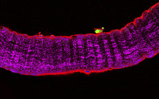
As a cell biologist, I dealt with analytical cytology quite early in my career. Whilst not being exclusively specialized in flow cytometry, I exploit the incredible power and the elegant performance of flow cytometry analysis to address several questions related to my scientific interests. I am mostly interested in the control of skeletal muscle differentiation and homeostasis and, more recently, in tissue engineering applications for regenerative medicine of this tissue. I had the honor to become an ISAC Scholar in 2006 and since them I am member of this society. Interests and vision I came across ISAC through my mentor, Dr. Laura Teodori at a time when I was doing my postdoctoral training about 10 years ago. Being scientifically seduced by the powerful applications of analytical cytology I started to attend ISAC international congresses and to participate more actively to the initiatives of the Society. At the XXIII ISAC congress in Quebec in 2006, I was awarded the ISAC Scholarship. As an ISAC Scholar I was encouraged to collaborate to the educational and organization strategies of the Society. I was young, mobile and without a tenured position. I felt sympathetic with the younger members of the Society and concerned about the typical issues they have to deal with: practicing, traveling, finding the resources to do that. By co-chairing a subcommittee of the MSC (Membership Services Committee) dedicated to Students' services in 2004-'08, I helped with the divulgation of skills and resources aimed to improve student members' success rate when applying to mobility grants. For instance, at the XXIV ISAC congress in Budapest in 2008 I participated to the Scientific Professional Skills Workshop with a presentation entitled “Short term mobility grants: tips and hints.” These issues found concrete development in the ISAC web page highlighting grants opportunities for short term mobility and for young fellows (originally published on the ISAC web site, http://www.isac-net.org/content/view/693/137/). I was responsible for that page, updating it twice a year and offering tutoring and advice to ISAC student members, with the aim to help their mobility and grant rising capacity. Today, from a more mature position inside the Society I wish to contribute to consolidate ISAC strengths and to further develop its potentials, hence my interest in the candidature for an ISAC Councilor position. In this position I could possibly exploit my growing experience and creative attitude to serve our common goals. I foresee two critical issues ISAC shall deal with in the incoming years: 1) geographical and intergenerational growth and 2) interaction with novel scientific and technological research areas. 1) I am convinced that our Society should invest more than ever on youngs. As far as I know ISAC educational and scholarship programs see an unprecedented success, which highlights the interests into our society by young researchers. ISAC should be even more attractive than today for them. In order to do so, we should pursue our politic of open access for young members, and of tutoring and education initiatives. Also, ISAC could set up initiatives aimed to assist its younger members in fund rising (startups, mobility). The initiatives could range from helping members to find senior partners for big grant applications to assisting members to identify calls and apply to them (a task often performed by specific services that are present only in major departments and universities). Obviously, the current programs dedicated to tutoring and visiting initiatives for young members would be synergistic with the novel actions I propose. An international society such as ISAC should become the catalyzer for exchanges and interactions not only vertically (between generations) but also horizontally (between emerging countries, where it is largely underrepresented, and consolidated scientific environments). So I would love to see novel initiatives, aimed at networking and diffusing the analytical cytology, especially targeted to the younger members and to researchers from emerging countries. 2) While being well developed in the US, tissue engineering and regenerative medicine are novel, fast growing disciplines in several countries, including european countries such as Italy and France, or Asian giants such as India and China. This area of Medicine is attracting more and more public and private financing, given its translational nature and high technological content, which in turns stimulates a growing involvement by scientists. Given the foreseen rapid shift to clinical practice in this field (indeed a reality for certain applications) it is of pivotal importance to set up at the same time innovative approaches and safety/quality control procedures for stem cell isolation and transfer, as well as for immunological stereotyping of host-implant interactions. In this context there is an important opportunity for ISAC to become the reference for such procedures and approaches. Thus, I would like to establish initiatives aimed to boost the collaboration between ISAC and non-ISAC members for regenerative medicine applications. An other level of interaction could be within the journals associated to those societies which represent the analogues of our Cytometry; politics encouraging cross-publication (and even cross-advertising for the scientific societies) could be built and I would be happy to collaborate on this. I wish that the publication of this text can be a matter of discussion and engagement by others and myself independently from my candidature as ISAC councilor. I really think that these issues are relevant for ISAC development and I am looking forward to seeing them dealt with.

2/19/2012

Quencing autofluorescence

Method for quencing background fluorescence due to aldehydes or autofluorescence

THE NETWORK OF OUR COLLABORATORS 2017

THE NETWORK OF OUR COLLABORATORS 2017
We collaborate with the Myology Group and the Cochin Hospital in Paris for stem cell studies and SRF, with the Cancer Centre at Ohio State University, Columbus for studies on the mechanisms underlying cachexia, with the Neurorehabilitation Unit at University of Pisa for clinical studies, with Pharmacology and Bioinformatics at the University of Urbino for advanced statistical analyses, with the Anatomy Section at the University of Perugia and with GYN/OB at the University of Western Piedmont for studies related to circulating factors and myogenic cell responses in cachexia, with the Biotech-Med Unit at ENEA, Chemistry in Rome and Anatomy in palermo for tissue engineering applications. Functional studies are carried out in our Departement in Rome in collaboration with Musaro's laboratory.