
Cancer cachexia
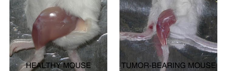
Compared to a control mouse (left) a tumor-bearing mouse (right) displays a dramatic muscle wasting. This loss of muscle mass is called cancer cachexia.
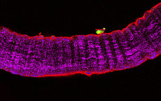
Exogenous gene expression in regenerating muscle
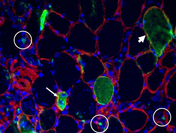
Depicted here is the over-expression of Green Fluorescent Protein (GFP, green; click on the image to access Tsien's Lab) in interstitial cells (circled), nascent myofibers (arrow) and adult fibers (arrowhead), in a regenerating Tibialis Anterior following focal injury. Laminin staining (red) highlights the basement membrane surrounding the skeletal muscle tissue, while nuclei are stained in blue. We do gene delivery by electroporation to study the regulation of muscle regeneration.
Cultures of myotubes on a conductive surface in a parallel orientation.
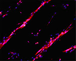
C2C12 cells cultured on gold, by mean of adhesion to 100 nm-wide stripes coated with anti Stem Cell antigen1 (Sca1) Ab. Nuclei (blue) and actin cytoskeleton (red) staining highlights the selective cells adhesion on the Ab-coated stripes and the formation of parallel multinucleated syncytia (myotubes).
9/16/2014
Everything you always wanted to know about SYNEMIN and never dared to ask
Synemin, a type IV intermediate filament (IF) protein, forms a bridge between IFs and cellular membrane, by interacting with Desmin, Vinculin, Talin and Dystrophin. An A-kinase anchoring protein, it also provides temporal and spatial targeting of protein kinase A. This protein likely functions to integrate mechanical stress and maintain structural integrity and tissue homeostasis in skeletal muscle and other cells.
 In our recent article, entitled "Synemin acts as a regulator of signalling molecules during skeletal muscle hypertrophy" (Li et al. J Cell Sci, 2014) we provide novel evidence on its role by phenotyping the synemin null mice.
All the additional hints and informations on synemin role in development and postnatal life in PubMed.
In our recent article, entitled "Synemin acts as a regulator of signalling molecules during skeletal muscle hypertrophy" (Li et al. J Cell Sci, 2014) we provide novel evidence on its role by phenotyping the synemin null mice.
All the additional hints and informations on synemin role in development and postnatal life in PubMed.
9/09/2014
Inflammation in Muscle Repair, Aging, and Myopathies
I have recently had the honor to co-edit a special issue of BioMed Research International dedicated to the role of inflammation in various acute and chronic conditions of muscle loss and disease.
Our group in Rome participated to the special issue with a paper (from which the figure is extracted) confirming in vivo a role for the neurohypophyseal hormone vasopressin in muscle repair and homeostasis. The paper by Alessandra Costa et al., entitled "Local Overexpression of V1a-Vasopressin Receptor Enhances Regeneration in Tumor Necrosis Factor-Induced Muscle Atrophy" can be found by fillowing the link.
 Figure legend. V1aR overexpression counteracts TNF-dependent protein degradation by stimulating the Akt pathway. (a) Western blots of phosphorylated Akt and native and phosphorylated FoxO3a expression demonstrate that in muscle overexpressing TNF, phospho-Akt and phospho-FoxO3a are downregulated, while the native Foxo3a is increased. In V1aR overexpressing muscles, the expression levels of phospho-FoxO3a and phospho-Akt is increased compared with TNF alone, while the native Foxo3a is reduced. (b–d) Densitometric analysis of three independent experiments of phospho-Akt, phospho-FoxO3a, and native FoxO3a expression levels. (e) Real-time PCR analysis revealed that the strong upregulation of atrogin-1 expression observed in the sample overexpressing TNF alone is downregulated in V1aR+TNF-transfected muscles. ; by Student’s -test.
 Here is the Table of content of the special issue, a nice mix of original research and review articles.
Inflammation in Muscle Repair, Aging, and Myopathies
Guest Editors: Marina Bouché, Pura Muñoz-Cánoves, Fabio Rossi, and Dario Coletti
Inflammation in Muscle Repair, Aging, and Myopathies, Marina Bouché, Pura Muñoz-Cánoves, Fabio Rossi, and Dario Coletti
Volume 2014 (2014), Article ID 821950, 3 pages
Stem Cell Transplantation for Muscular Dystrophy: The Challenge of Immune Response, Sara Martina Maffioletti, Maddalena Noviello, Karen English, and Francesco Saverio Tedesco
Volume 2014 (2014), Article ID 964010, 12 pages
From Innate to Adaptive Immune Response in Muscular Dystrophies and Skeletal Muscle Regeneration: The Role of Lymphocytes, Luca Madaro and Marina Bouché
Volume 2014 (2014), Article ID 438675, 12 pages
Cardioprotective Effects of Osteopontin-1 during Development of Murine Ischemic Cardiomyopathy, Georg D. Duerr, Bettina Mesenholl, Jan C. Heinemann, Martin Zoerlein, Peter Huebener, Prisca Schneider, Andreas Feisst, Alexander Ghanem, Klaus Tiemann, Daniela Dewald, Armin Welz, and Oliver Dewald
Volume 2014 (2014), Article ID 124063, 15 pages
IL-6 Impairs Myogenic Differentiation by Downmodulation of p90RSK/eEF2 and mTOR/p70S6K Axes, without Affecting AKT Activity, Michele Pelosi, Manuela De Rossi, Laura Barberi, and Antonio Musarò
Volume 2014 (2014), Article ID 206026, 12 pages
Local Overexpression of V1a-Vasopressin Receptor Enhances Regeneration in Tumor Necrosis Factor-Induced Muscle Atrophy, Alessandra Costa, Angelica Toschi, Ivana Murfuni, Laura Pelosi, Gigliola Sica, Sergio Adamo, and Bianca Maria Scicchitano
Volume 2014 (2014), Article ID 235426, 14 pages
Influence of Immune Responses in Gene/Stem Cell Therapies for Muscular Dystrophies, Andrea Farini, Clementina Sitzia, Silvia Erratico, Mirella Meregalli, and Yvan Torrente
Volume 2014 (2014), Article ID 818107, 16 pages
Vitamin D Receptor Agonists: Suitable Candidates as Novel Therapeutic Options in Autoimmune Inflammatory Myopathy, Clara Crescioli
Volume 2014 (2014), Article ID 949730, 10 pages
7-Tesla Magnetic Resonance Imaging Precisely and Noninvasively Reflects Inflammation and Remodeling of the Skeletal Muscle in a Mouse Model of Antisynthetase Syndrome, Clara Sciorati, Antonio Esposito, Lara Campana, Tamara Canu, Antonella Monno, Anna Palmisano, Francesco De Cobelli, Alessandro Del Maschio, Dana P. Ascheman, Angelo A. Manfredi, and Patrizia Rovere-Querini
Volume 2014 (2014), Article ID 879703, 8 pages
Understanding the Process of Fibrosis in Duchenne Muscular Dystrophy, Yacine Kharraz, Joana Guerra, Patrizia Pessina, Antonio L. Serrano, and Pura Muñoz-Cánoves
Volume 2014 (2014), Article ID 965631, 11 pages
Inflammation Based Regulation of Cancer Cachexia, Jill K. Onesti and Denis C. Guttridge
Volume 2014 (2014), Article ID 168407, 7 pages
Macrophage Plasticity in Skeletal Muscle Repair, Elena Rigamonti, Paola Zordan, Clara Sciorati, Patrizia Rovere-Querini, and Silvia Brunelli
Volume 2014 (2014), Article ID 560629, 9 pages
Here is the Table of content of the special issue, a nice mix of original research and review articles.
Inflammation in Muscle Repair, Aging, and Myopathies
Guest Editors: Marina Bouché, Pura Muñoz-Cánoves, Fabio Rossi, and Dario Coletti
Inflammation in Muscle Repair, Aging, and Myopathies, Marina Bouché, Pura Muñoz-Cánoves, Fabio Rossi, and Dario Coletti
Volume 2014 (2014), Article ID 821950, 3 pages
Stem Cell Transplantation for Muscular Dystrophy: The Challenge of Immune Response, Sara Martina Maffioletti, Maddalena Noviello, Karen English, and Francesco Saverio Tedesco
Volume 2014 (2014), Article ID 964010, 12 pages
From Innate to Adaptive Immune Response in Muscular Dystrophies and Skeletal Muscle Regeneration: The Role of Lymphocytes, Luca Madaro and Marina Bouché
Volume 2014 (2014), Article ID 438675, 12 pages
Cardioprotective Effects of Osteopontin-1 during Development of Murine Ischemic Cardiomyopathy, Georg D. Duerr, Bettina Mesenholl, Jan C. Heinemann, Martin Zoerlein, Peter Huebener, Prisca Schneider, Andreas Feisst, Alexander Ghanem, Klaus Tiemann, Daniela Dewald, Armin Welz, and Oliver Dewald
Volume 2014 (2014), Article ID 124063, 15 pages
IL-6 Impairs Myogenic Differentiation by Downmodulation of p90RSK/eEF2 and mTOR/p70S6K Axes, without Affecting AKT Activity, Michele Pelosi, Manuela De Rossi, Laura Barberi, and Antonio Musarò
Volume 2014 (2014), Article ID 206026, 12 pages
Local Overexpression of V1a-Vasopressin Receptor Enhances Regeneration in Tumor Necrosis Factor-Induced Muscle Atrophy, Alessandra Costa, Angelica Toschi, Ivana Murfuni, Laura Pelosi, Gigliola Sica, Sergio Adamo, and Bianca Maria Scicchitano
Volume 2014 (2014), Article ID 235426, 14 pages
Influence of Immune Responses in Gene/Stem Cell Therapies for Muscular Dystrophies, Andrea Farini, Clementina Sitzia, Silvia Erratico, Mirella Meregalli, and Yvan Torrente
Volume 2014 (2014), Article ID 818107, 16 pages
Vitamin D Receptor Agonists: Suitable Candidates as Novel Therapeutic Options in Autoimmune Inflammatory Myopathy, Clara Crescioli
Volume 2014 (2014), Article ID 949730, 10 pages
7-Tesla Magnetic Resonance Imaging Precisely and Noninvasively Reflects Inflammation and Remodeling of the Skeletal Muscle in a Mouse Model of Antisynthetase Syndrome, Clara Sciorati, Antonio Esposito, Lara Campana, Tamara Canu, Antonella Monno, Anna Palmisano, Francesco De Cobelli, Alessandro Del Maschio, Dana P. Ascheman, Angelo A. Manfredi, and Patrizia Rovere-Querini
Volume 2014 (2014), Article ID 879703, 8 pages
Understanding the Process of Fibrosis in Duchenne Muscular Dystrophy, Yacine Kharraz, Joana Guerra, Patrizia Pessina, Antonio L. Serrano, and Pura Muñoz-Cánoves
Volume 2014 (2014), Article ID 965631, 11 pages
Inflammation Based Regulation of Cancer Cachexia, Jill K. Onesti and Denis C. Guttridge
Volume 2014 (2014), Article ID 168407, 7 pages
Macrophage Plasticity in Skeletal Muscle Repair, Elena Rigamonti, Paola Zordan, Clara Sciorati, Patrizia Rovere-Querini, and Silvia Brunelli
Volume 2014 (2014), Article ID 560629, 9 pages
Our editorial "Inflammation in Muscle Repair, Aging, and Myopathies" briefly summarizes the main focus of this special issue, i.e. bringing together studies that used different experimental approaches in vivo or in vitro to dissect the dynamic changes taking place in specific immune cell populations, their cross talk with other cell types within the muscle milieu, and their contribution to normal versus pathological muscle repair. While the number of scientific publications on the topic of skeletal muscle inflammation has steadily grown over the last two decades, the notion of inflammation as a common feature in muscle degeneration occurring in aging and myopathies and its association with altered muscle has to our knowledge never previously been addressed and discussed in dedicated journal issues before.

Subscribe to:
Posts (Atom)
THE NETWORK OF OUR COLLABORATORS 2017
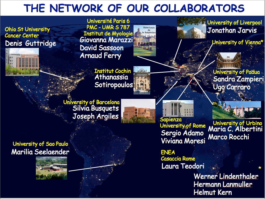
We collaborate with the Myology Group and the Cochin Hospital in Paris for stem cell studies and SRF, with the Cancer Centre at Ohio State University, Columbus for studies on the mechanisms underlying cachexia, with the Neurorehabilitation Unit at University of Pisa for clinical studies, with Pharmacology and Bioinformatics at the University of Urbino for advanced statistical analyses, with the Anatomy Section at the University of Perugia and with GYN/OB at the University of Western Piedmont for studies related to circulating factors and myogenic cell responses in cachexia, with the Biotech-Med Unit at ENEA, Chemistry in Rome and Anatomy in palermo for tissue engineering applications. Functional studies are carried out in our Departement in Rome in collaboration with Musaro's laboratory.


