RESEARCH INTERESTS: Cellular and molecular mechanisms of striated muscle physiopathology
1. PHARMACOLOGICAL, PHYSICAL, AND NUTRITIONAL INTERVENTIONS AGANIST CANCER CACHEXIA: My laboratory is focused on different approaches to counteract cancer cachexia, including pharmacological (exercise mimetics), physiological (physical activity), and nutritional (supplements) interventions in humans and animal models.
2. MYOFIBER MEMBRANE DAMAGE AND REPAIR: Duchenne Muscular Dystrophy (DMD), is a lethal genetic, muscle-wasting disease, characterized by progressive muscle fragility and weakness. The muscle membrane repair mechanism (MRM) is an active resealing pathway involving vesicle-sarcolem fusion to “patch” the compromised plasma membrane and represents a possible target to counteract muscle wasting in DMD, in which the chronic cycle of muscle degeneration-regeneration plays a pivotal role in disease progression.
3. PATENTS AND TECHNOLOGY TRASNFER: I am co-inventor of a patented procedure to produce Hsp60-enriched exosomes with exercise-mimetic activity, a product that is, therefore, called Physiactisome. Patent: Physiactisome – «Procedure for the synthesis of HSP-containing exosomes and their use against muscle atrophy and cachexia» - patent n. 102018000009235 on 8/10/2018, deposited by Università di Palermo. Owners: Università di Palermo, Università di Roma La Sapienza, Nanovector Torino, Sorbonne Université. List of inventors: Valentina Di Felice, Rosario Barone, Antonella Marino Gammazza, Campanella Claudia, Cappello Francesco, Farina Felicia, Eleonora Trovato, Daniela D’Amico, Filippo Macaluso, Dario Coletti, Sergio Adamo, Gabriele Multhoff, Paolo Gasco. International publication number WO 2020/075004 A1. This product can be exploited against muscle atrophy, since it ameliorates muscle endurance and homeostasis. The presentation of the product and the corresponding Spinoff project (iBioTHEx) was awarded the third prize at the EIT JumpStarter Grand final, Riga, Latvia, 15-17/11/2019, Health category.
4. PHYSIOPATHOLOGY OF MUSCLE TISSUES: I contribute to discovering and explaining those mechanisms underlying pathologies of the striated and smooth muscle tissues; this activity is carried out at Sorbonne University by using genetic murine models.
RESEARCH INTERESTS: Tissue engineering of skeletal muscle
Background and rationale.
Tissue engineering lies at the interface of regenerative medicine and developmental biology, and represent an innovative and multidisciplinary approach to build organs and tissues (Ingber and Levin, Development 2007). The skeletal muscle is a contractile tissue characterized by highly oriented bundles of giant syncytial cells (myofibers) and by mechanical resistance. Contractile, tissue-engineered skeletal muscle would be of significant benefit to patients with muscle deficits secondary to congenital anomalies, trauma, or surgery. Obvious limitations to this approach are the complexity of the musculature, composed of multiple tissues intimately intermingled and functionally interconnected, and the big dimensions of the majority of the muscles, which imply the involvement of an enormous amount of cells and rises problems of cell growth and survival (nutrition and oxygen delivery etc.). Two major approaches are followed to address these issues. Self-assembled skeletal muscle constructs are produced in vitro by delaminating sheets of cocultured myoblasts and fibroblasts, which results in contractile cylindrical “myooids.” Matrix-based approaches include placing cells into compacted lattices, seeding cells onto degradable polyglycolic acid sponges, seeding cells onto acellularized whole muscles, seeding cells into hydrogels, and seeding nonbiodegradable fiber sheets. Recently, decellularized matrix from cadaveric organs has been proven to be a good scaffold for cell repopulation to generate functional hearts in mice (Ott et al. Nature Medicine 2008).
I have obtained cultures of skeletal muscle cells on conductive surfaces, which is required to develop electronic device–muscle junctions for tissue engineering and medical applications1. I aim to exploit this system for either recording or stimulation of muscle cell biological activities, by exploiting the field effect transistor and capacitor potential of the conductive substratum-cell interface. Also, we are able to create patterned dispositions of molecules and cells on gold, which is important to mimic the highly oriented pattern myofibers show in vivo.
I have found that Static magnetic fields enhance skeletal muscle differentiation in vitro by improving myoblast alignment2. Static magnetic field (SMF) interacts with mammal skeletal muscle; however, SMF effects on skeletal muscle cells are poorly investigated. 80 +/- mT SMF generated by a custom-made magnet promotes myogenic cell differentiation and hypertrophy in vitro. Finally, we have transplanted acellular scaffolds to study the in vivo response to this biomaterial3, which we want to exploit for tissue culture and regenerative medicine of skeletal muscle.
The specific aims of my current research are:
1) to increase and optimize the production and alignment of myogenic cells and myotubes in vitro;
2) to manipulate the niche of muscle stem cells aimed at ameliorating their regenerative capacity in vivo;
3) to develop muscle-electrical devices interactions. We plan to exploit the cell culture system on conductive substrates for either recording or stimulation of muscle cell biological activities, by exploiting the field effect transistor and capacitor potential of the conductive substratum-cell interface.
4) to better clarify the biological effects of Static Magnetic Fields. With the aim to characterize the molecular mechanism underlying the effects of SMF on cell differentiation and alignment we are exposing molecules and cells to SMF below 1T.
5) to produce pre-assembled, off-the-shelf skeletal muscle. We are seeding acellularized muscle scaffold with various cell types, with the goal to obtain functional muscle with vascular supply and nerves.
REFERENCES
1) Coletti D. et al., J Biomed Mat Res 2009; 91(2):370-377.
2) Coletti D. et al., Cytometry A. 2007;71(10):846-56.
3) Perniconi B. et al. Biomaterials, 2011 in press

Figure legend. Examples of focal injuries. (LEFT) Hematoxilin-and eosin-stained murine skeletal muscle, longitudinally sectioned to show the gaps in three adjacent fibers. The injury likely occurred following an intense exercise session (wheel running). Upon leakage of the broken sarcolemma, factors such as Wnt are released before a fast repair process known as patch repair occurs. In turn, Wnt factors trigger the activation of satellite cells and other resident interstitial cells with myogenic potential, which proliferate, migrate and fuse into small myotubes that ultimately fuse with the damaged fibers. (RIGHT) Toluidine blue-stained semithin section of a murine carotid showing damage, likely due to smooth muscle cell-restricted inactivation of the serum response factor gene. A rupture of the endothelial layer, as well as of the elastin matrix, with exposure of underlying cells is visible; release of intracellular factors (von Willebrand Factor) and exposure of undisclosed antigens (collagen) are essential for the subsequent phases of clot formation, remodeling and repair of the wall defect. Bar = 25 micron.

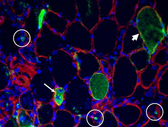
Figure legend. Macrophages infiltrate necrotic muscle fibers. Serial section of murine skeletal muscle in an area of necrosis; corresponding fibers are highlighted by matching color arrows. Evans Blue Dye (EBD) highlights muscle fibers whose plasma membrane is leaking owing to damage; hematoxylin and eosin staining (H&E), showing cellular infiltration in EBD + fibers; histochemistry for esterase staining highlights esterase-enriched macrophages; immunofluorescence analysis for activated macrophages expressing F4/80 (green) confirms the invasion of the muscle fibers by macropohages: laminin (red) and nuclei (blue) are also shown.
Coletti et al. Regenerative Medicine Research 2013 1:4 doi:10.1186/2050-490X-1-4

In this review article we present and compare the cellular aspects of regeneration in skin, nerve and muscle, three organs characterized by differences not only in anatomical and functional organization, but also in the number and location of stem cell niches and populations, which ultimately result in varying regenerative potential. By discussing the common traits and the specific features of regeneration in three model tissues, we propose general models of regeneration and highlight various strategies adopted to cope with damage and repair in mammals.The strategies used by a wide range of tissues to replace their lost parts vary, probably as result of evolutionary-based mechanisms for specific tissue regeneration. While the epidermis regenerates ex novo, neurons restore their missing parts; muscle fibers instead use a mixed strategy, based on the reconstruction of missing parts and on the generation of new fibers. These differential strategies are represented by the two terms used in the title to refer to different forms of regeneration: restoration, the attempt to re-establish the status quo ante, and reconstruction, a more radical response, characterized by ex novo cell colonization and tissue formation. The choice of either strategy is deeply influenced by the anatomy and the distribution/features of stem cell niches typical of a given organ. In addition, the energetic costs for either regenerative strategy are also likely to play an important role. The abstraction of divergences and analogies between different types of tissue regeneration might pave the way for the mathematical modeling of this process, thereby making a major contribution to both pathology and regenerative medicine.
When talking of wound healing, a distinction is made by some authors between regeneration and repair. Regeneration is used to refer to the complete replacement of damaged tissue with new tissue not associated with scar tissue, while repair is used to refer to the re-establishment of tissue continuity [1]. Regeneration can be attained by two means: a) restoration, defined as “putting together what is broken”; b) reconstruction, defined as “replacing and rebuilding what is torn down” (according to the Merriam-Webster Dictionary). To grant homeostasis, most tissues undergo continuous or cyclic processes of regeneration. Which of the afore-mentioned strategies tissues adopt depends on the histological features discussed in the article entitled "
Restoration versus reconstruction: cellular mechanisms of skin, nerve and muscle regeneration compared".


No comments:
Post a Comment