RESEARCH INTERESTS: Cellular and molecular mechanisms of striated muscle physiopathology
1. PHARMACOLOGICAL, PHYSICAL, AND NUTRITIONAL INTERVENTIONS AGANIST CANCER CACHEXIA: My laboratory is focused on different approaches to counteract cancer cachexia, including pharmacological (exercise mimetics), physiological (physical activity), and nutritional (supplements) interventions in humans and animal models.
2. MYOFIBER MEMBRANE DAMAGE AND REPAIR: Duchenne Muscular Dystrophy (DMD), is a lethal genetic, muscle-wasting disease, characterized by progressive muscle fragility and weakness. The muscle membrane repair mechanism (MRM) is an active resealing pathway involving vesicle-sarcolem fusion to “patch” the compromised plasma membrane and represents a possible target to counteract muscle wasting in DMD, in which the chronic cycle of muscle degeneration-regeneration plays a pivotal role in disease progression.
3. PATENTS AND TECHNOLOGY TRASNFER: I am co-inventor of a patented procedure to produce Hsp60-enriched exosomes with exercise-mimetic activity, a product that is, therefore, called Physiactisome. Patent: Physiactisome – «Procedure for the synthesis of HSP-containing exosomes and their use against muscle atrophy and cachexia» - patent n. 102018000009235 on 8/10/2018, deposited by Università di Palermo. Owners: Università di Palermo, Università di Roma La Sapienza, Nanovector Torino, Sorbonne Université. List of inventors: Valentina Di Felice, Rosario Barone, Antonella Marino Gammazza, Campanella Claudia, Cappello Francesco, Farina Felicia, Eleonora Trovato, Daniela D’Amico, Filippo Macaluso, Dario Coletti, Sergio Adamo, Gabriele Multhoff, Paolo Gasco. International publication number WO 2020/075004 A1. This product can be exploited against muscle atrophy, since it ameliorates muscle endurance and homeostasis. The presentation of the product and the corresponding Spinoff project (iBioTHEx) was awarded the third prize at the EIT JumpStarter Grand final, Riga, Latvia, 15-17/11/2019, Health category.
4. PHYSIOPATHOLOGY OF MUSCLE TISSUES: I contribute to discovering and explaining those mechanisms underlying pathologies of the striated and smooth muscle tissues; this activity is carried out at Sorbonne University by using genetic murine models.
RESEARCH INTERESTS: Tissue engineering of skeletal muscle
Background and rationale.
Tissue engineering lies at the interface of regenerative medicine and developmental biology, and represent an innovative and multidisciplinary approach to build organs and tissues (Ingber and Levin, Development 2007). The skeletal muscle is a contractile tissue characterized by highly oriented bundles of giant syncytial cells (myofibers) and by mechanical resistance. Contractile, tissue-engineered skeletal muscle would be of significant benefit to patients with muscle deficits secondary to congenital anomalies, trauma, or surgery. Obvious limitations to this approach are the complexity of the musculature, composed of multiple tissues intimately intermingled and functionally interconnected, and the big dimensions of the majority of the muscles, which imply the involvement of an enormous amount of cells and rises problems of cell growth and survival (nutrition and oxygen delivery etc.). Two major approaches are followed to address these issues. Self-assembled skeletal muscle constructs are produced in vitro by delaminating sheets of cocultured myoblasts and fibroblasts, which results in contractile cylindrical “myooids.” Matrix-based approaches include placing cells into compacted lattices, seeding cells onto degradable polyglycolic acid sponges, seeding cells onto acellularized whole muscles, seeding cells into hydrogels, and seeding nonbiodegradable fiber sheets. Recently, decellularized matrix from cadaveric organs has been proven to be a good scaffold for cell repopulation to generate functional hearts in mice (Ott et al. Nature Medicine 2008).
I have obtained cultures of skeletal muscle cells on conductive surfaces, which is required to develop electronic device–muscle junctions for tissue engineering and medical applications1. I aim to exploit this system for either recording or stimulation of muscle cell biological activities, by exploiting the field effect transistor and capacitor potential of the conductive substratum-cell interface. Also, we are able to create patterned dispositions of molecules and cells on gold, which is important to mimic the highly oriented pattern myofibers show in vivo.
I have found that Static magnetic fields enhance skeletal muscle differentiation in vitro by improving myoblast alignment2. Static magnetic field (SMF) interacts with mammal skeletal muscle; however, SMF effects on skeletal muscle cells are poorly investigated. 80 +/- mT SMF generated by a custom-made magnet promotes myogenic cell differentiation and hypertrophy in vitro. Finally, we have transplanted acellular scaffolds to study the in vivo response to this biomaterial3, which we want to exploit for tissue culture and regenerative medicine of skeletal muscle.
The specific aims of my current research are:
1) to increase and optimize the production and alignment of myogenic cells and myotubes in vitro;
2) to manipulate the niche of muscle stem cells aimed at ameliorating their regenerative capacity in vivo;
3) to develop muscle-electrical devices interactions. We plan to exploit the cell culture system on conductive substrates for either recording or stimulation of muscle cell biological activities, by exploiting the field effect transistor and capacitor potential of the conductive substratum-cell interface.
4) to better clarify the biological effects of Static Magnetic Fields. With the aim to characterize the molecular mechanism underlying the effects of SMF on cell differentiation and alignment we are exposing molecules and cells to SMF below 1T.
5) to produce pre-assembled, off-the-shelf skeletal muscle. We are seeding acellularized muscle scaffold with various cell types, with the goal to obtain functional muscle with vascular supply and nerves.
REFERENCES
1) Coletti D. et al., J Biomed Mat Res 2009; 91(2):370-377.
2) Coletti D. et al., Cytometry A. 2007;71(10):846-56.
3) Perniconi B. et al. Biomaterials, 2011 in press
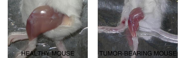
We have recently shown (
Pigna et al., link to full text) in mice that the striking beneficial effects exerted by endurance exercise (running) on survival and muscle wasting (cancer cachexia) are due to a rescue of the autophagic flux, which is altered in pathological conditions such as cachexia. This counterintuitive observation (autophagy means "to eat onself" and it is a way to grant cell survival in the absence of nutrients, accounting for cell atrophy) was done on both tumor-bearing humans and mice. In addition to the proposed molecular mechanisms underlying exercise effects, we showed that exercise can be replaced by pharmacological treatments, such as rapamycin or AICAR (5-aminoimidazole-4-carboxamide-1-beta-D-ribofuranoside), the latter also known as exercise mimetic (EM). EM are a heterogeneous group of compounds that share the ability to induce pathways which are physiologically activated by exercise, thus stimulating endurance and rescuing muscle atrophy. GW1516 (also known as GW501516) or AICAR, among others, are activators of AMPK, PPARs and PGC-1. This pathway ultimately leads to the activation of both mitochondriogenesis and muscle oxidative metabolism, as it would in response to an increased AMP/ATP ratio, physiologically following exercise and energy consumption. As enhancers of physical performance, EM treatments would be considered doping agents in sport.However, thay couls be to avoid many consequences of inactivity due to aging, reduced gravity and forced immobilization; moreover, it could be prescribed for severely diseased people, such as cancer patients, for whom doing exercise could not be a streightforward endevour.
Further discussion on the new view of the exercised muscle as an endocrine organ and the promise of exercise mimetics can be found in our editorial entitled "
Will exercise mimetics hold promise?"

Dietary bioactive compounds singificanlty affect physiology and pathology in our lives. This is not just an indirerct effect, due to lower risk of odd pathological conditions deriving from food intake, such as the link between colesterol intake and stroke. What I mean is that fatty acids biosynthesis and metabolism may have direct effects on molecular events of pivotal importance for healthy life, such as DNA damage response (DDR), which, in turn, is implicate din cancer and aging.
We have recently done examined the influence of dietary bioactive compounds on the micro-RNAs (miRs) involved in DDR, which potentially may offer clues on the role of these compounds as potential chemopreventive and antiaging (geroprotective) modalities. The results of our analysis (
link to full text here) show a strong impact of the compounds considered on important miRs involved in DD/DDR pathways. Epi-gallocatechin-3-gallate (EGCG), resveratrol (RSV), curcumin (CRC) and n3-PUFA (n3-polyunsaturated fatty acids) are among the most studied compounds shown to have beneficial effects on human health. The analysis suggests a synergic action of all these compounds in the modulation of six miRs. Among these common miRs, we found miR-21 and miR-146. The results of the analysis of the pathways allowed us to speculate how food intervention could modulate DD/DDR.
Figure legend. Venn diagram showing the microRNAs involved in DD/DDR (ellipse, grey) and identified as modulated by bioactive compounds: EGCG (epi-gallocatechin-3-gallate; green), CRC (curcumin; blue), RSV (resveratrol; pink) and n3-PUFAs (n3-polyunsaturated fatty acids, yellow). The common miRNAs, modulated by all four compounds, are indicated in red (miR-16, miR-25, miR-21, miR-181a,miR-146b, miR-34a).
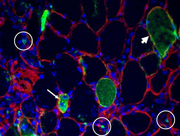
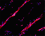
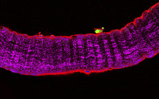
An experimental example of such beneficial effects exherted by the compounds above on a muscle pathology is shown in our study by Carotenuto et al. (
link to full text here) . In this paper we show that dietary flaxseed ameliorate skeletal muscle regeneration in an animal model of muscular dystrophy.Flaxseed is one of the richest sources of n-3 PUFA acid
α-linolenic acid (ALA). ALA has been shown to have many beneficial effects in several experimental and clinical setting by targeting critical players
involved in inflammation response, tissue regeneration and repair.Our study demonstrates that the pathological status of muscular dystrophy can be effectively mitigated by administering natural compounds contained in flaxseed that have a pleiotropic effect rather than by targeting a specific gene
mutation as occurs in other therapeutic strategies. Flaxseed not only repairs the damage in the sarcolemma, which is the primary consequence of gene mutation, but also exerts a more extensive epigenetic action that stimulates multiple molecular pathways in dystrophic muscles.

Figure legend. Flaxseed-enriched diet preserves dystrophic skeletal muscle morphology. For all in vivo observations, dystrophic hamsters were fed with a flaxseed- enriched diet (FS) from weaning to the age of 150 days (Dystr/FS) and compared with dystrophic (Dystr/P) and healthy hamsters (Healthy) fed with standard pellet (P). (A) Representative images of H&E-stained skeletal muscle sections. Scale bar: 50 μm. (B) Percentage of myofibers with internalized nuclei from H&E-stained skeletal muscle sections. *P<0.05 vs. Healthy; § P<0.05 vs. Dystr/P; n=5. (C) Component Score Coefficient Matrix. The coefficients by which variables are multiplied to obtain factor scores are shown. The variables are represented by the morphometric parameters derived from light microscope images of skeletal muscle (six sections from each of 5 animals/group). The values highlighted (red boxes) indicate the variables most closely associated with Principal Components 1 and 2. (D) Principal Component Analysis (PCA). Three series of data from of Healthy, Dystr/FS and Dystr/P hamsters were plotted in the bidimensional space defined by the 1st and 2nd PCA. FS diet (Dystr/FS, green dots) restored the morphological parameters of the dystrophic (Dystr/P, yellow dots) phenotype towards value closer to those of healthy (blue dots) skeletal muscles.
In addition to the all above, linolenic acid seems to protect muscle cells from cytokine-induced apoptosis, as shown by us in the paper Cartoenuto et al. (
link to full text here); this could singificantly contribute to promote survival in disease states characterized by chronic inflammation due to muscle damage, such as muscle dystrophy or cachexia.


