
Cancer cachexia
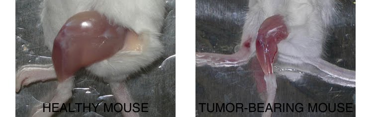
Compared to a control mouse (left) a tumor-bearing mouse (right) displays a dramatic muscle wasting. This loss of muscle mass is called cancer cachexia.
Exogenous gene expression in regenerating muscle
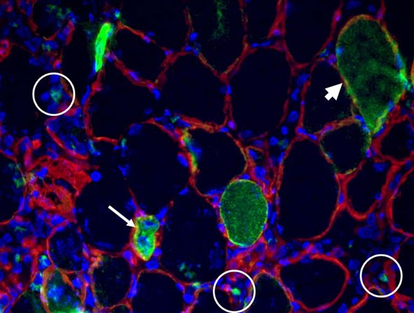
Depicted here is the over-expression of Green Fluorescent Protein (GFP, green; click on the image to access Tsien's Lab) in interstitial cells (circled), nascent myofibers (arrow) and adult fibers (arrowhead), in a regenerating Tibialis Anterior following focal injury. Laminin staining (red) highlights the basement membrane surrounding the skeletal muscle tissue, while nuclei are stained in blue. We do gene delivery by electroporation to study the regulation of muscle regeneration.
Cultures of myotubes on a conductive surface in a parallel orientation.
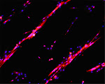
C2C12 cells cultured on gold, by mean of adhesion to 100 nm-wide stripes coated with anti Stem Cell antigen1 (Sca1) Ab. Nuclei (blue) and actin cytoskeleton (red) staining highlights the selective cells adhesion on the Ab-coated stripes and the formation of parallel multinucleated syncytia (myotubes).
9/25/2015
Focus: Biomaterials and bioactive molecules to drive differentiation in striated muscle tissue
Below the point on our most recent research in Tissue enginnering.
Recently, an article by Armstrong et al. entitled « TOP 10 DEVELOPMENTS IN STEM CELL BIOLOGY OVER THE LAST 30 YEARS » summarized ten quantum leaps in stem cells research.
These are:
1) THE DISCOVERY AND ISOLATION ADULT STEM CELLS
2) THE FIRST EMBRYONIC STEM CELLS FROM MICE
3) THE DEVELOPMENT OF MAMMALIAN CLONING
4) HUMAN EMBRYONIC STEM CELLS
5) THE CONCEPT OF CANCER STEM CELLS
6) INDUCED PLURIPOTENT STEM CELLS
7) MESENCHYMAL STEM CELLS
8) THE TISSUE ENGINEERING WITH STEM CELLS
9) THE IMPROVEMENT OF GENETIC MANIPULATION
THE BEGINNING OF TRANSLATIONAL MEDICAL APPLICATIONS
(In: Armstrong L. et al. STEM CELLS 2012;30:2–9. link fo tull text: https://drive.google.com/file/d/0B_z0HQaQV25yX3EzOTVBcXNSVTA/view?usp=sharing)
We have cited in this article (Perniconi et al. Biomaterials, 2011) as an outstanding example of tissue engineering approach. That's great! Thank you.
We mainly focus on the use of Biomaterials to drive differentiation in striated muscle tissue. The latter has a peculiar way of regeneraitng (Restoration versus reconstruction: cellular mechanisms of skin, nerve and muscle regeneration compared. Coletti D, Teodori L, Lin Z, Beranudin JF, Adamo S. Regen Med Res. 2013 Oct 1;1(1):4. doi: 10.1186/2050-490X-1-4. eCollection 2013 Dec. Review. PMID: 25984323. Full text here).
Advances in tissue replacement and regeneraiton involve muscle replacement, such as in the case of volumetric muscle loss, and inflammation control, such as in myopathies characterized by chronic inflammation (Inflammation in muscle repair, aging, and myopathies. Bouché M, Muñoz-Cánoves P, Rossi F, Coletti D. Biomed Res Int. 2014;2014:821950. doi: 10.1155/2014/821950. Epub 2014 Aug 4. No abstract available. PMID: 25162030. Full text here).
Of the various alternative approaches to replace or regenerate muscle, the use of biomaterials has gained major attention (Biomaterials and bioactive molecules to drive differentiation in striated muscle tissue engineering. Di Felice V, Forte G, Coletti D. Front Physiol. 2015 Feb 23;6:52. doi: 10.3389/fphys.2015.00052. eCollection 2015. PMID: 25755644. Full text here).
In particular, scaffolds obtained by decellularization of small intestinal submucosa(SIS),urinary bladder mucosa(UB) and skeletal muscle are getting very promising results at pre-clinical and clinical level (reviewed in Native extracellular matrix: a new scaffolding platform for repair of damaged muscle. Teodori L, Costa A, Marzio R, Perniconi B, Coletti D, Adamo S, Gupta B, Tarnok A. Front Physiol. 2014 Jun 16;5:218. doi: 10.3389/fphys.2014.00218. eCollection 2014. Review. PMID: 24982637. Full text here). A consistent body of evidence indicates that extra-cellular matrix (ECM) proteins regulate stem cell differentiation and renewal and are highly relevant to tissue engineering applications. The ECM also provides a supportive medium for blood or lymphatic vessels and for nerves. Thus, the ECM is the nature's ideal biological scaffold material.
We have been focusing on ECM derived from decellularized skeletal muscle. This muscle acellular scaffold (MAS) may represent a suitable environment for muscle and non-muscle 3D constructs characterized by a highly organized structure whose relative stability promotes integration with the surrounding tissues. (Muscle acellular scaffold as a biomaterial: effects on C2C12 cell differentiation and interaction with the murine host environment. Perniconi B, Coletti D, Aulino P, Costa A, Aprile P, Santacroce L, Chiaravalloti E, Coquelin L, Chevallier N, Teodori L, Adamo S, Marrelli M, Tatullo M. Front Physiol. 2014 Sep 26;5:354. doi: 10.3389/fphys.2014.00354. eCollection 2014. PMID: 25309452. Full text here).
Our recent work also highlights the plasticity of MAS, suggesting that it may be possible to consider MAS for a wider range of tissue engineering applications than the mere replacement of volumetric muscle loss. (Muscle extracellular matrix scaffold is a multipotent environment. Aulino P, Costa A, Chiaravalloti E, Perniconi B, Adamo S, Coletti D, Marrelli M, Tatullo M, Teodori L. Int J Med Sci. 2015 Apr 6;12(4):336-40. doi: 10.7150/ijms.10761. eCollection 2015. PMID: 25897295. Full text here).
Nonetheless, caution is imposed when announcing these major progresses on skeletal muscle tissue engineering, since is still impossible to fully reconstruct such a highly hierarchized, big, and complex organ for in vivo transplantaiton. While for these ambitious in vivo tissue engineering applications, there may still be a long way to go, novel in vitro applications for tissue engineered contructs are emerging, such as 3D cultures aimed at better mimicking an in vivo models. It is self-evident that bidimensional cultures are very limited insofar as the physiological 3D tissue organization they yield is somewhat approximate. With the current need to develop experimentals models replacing or refining animal-based research, these ideas are becoming increasingly appealing. In conclusion, we believe that the best bet for skeletal muscle TE is to focus on specific, anatomically defined solutions or on 3D in vitro modeling of muscle tissue for basic and applied research (Skeletal muscle tissue engineering: best bet or black beast? Perniconi B, Coletti D. Front Physiol. 2014 Jul 4;5:255. doi: 10.3389/fphys.2014.00255. eCollection 2014. No abstract available. PMID: 25071600. Full text here). We are confident that we will eventually be able to transform the black beast (i.e., striated muscle tissue engineering) into the best bet (i.e., a successful clinical practice based on engineered muscles).
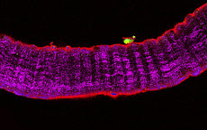
Figure from: Coletti Det al. Regen Med Res. 2013 Oct 1;1(1):4. Hematoxilin- and eosin-staining (H&E) and immunofluorescence
localization of muscle fiber damage (red) and of the membrane basement component laminin (green) on serial cross-sections of murine Tibialis anterior muscle (only a portion of
the muscle is shown). Thirty minutes before fixation, the muscle was subjected to two types of physical injury: mechanical stress by crunching
and tearing with forceps (LEFT) and freezing by applying a liquid nitrogen-cooled steel forceps to the surface (facing down in the picture) for
10 seconds (CENTER). Apart for the edema and fiber swelling visible in the images on the right, no major alterations of the basement membrane
are seen following focal injury. In mice injected with Evans Blue Dye (EBD, RIGHT), injury muscle fiber necrosis (red) is apparent 8 h after freezing
thanks to accumulation of EBD in the interior part of the damaged fibers. The muscle fibers die and are either renewed or replaced within the
intact scaffold represented by the membrane basement, which wraps each fiber.Note that in all cases the basement membrane remains intact in sharp contrast with muscle fiber damage, therefore the general architecture of the muscle is preserved.
Subscribe to:
Posts (Atom)
THE NETWORK OF OUR COLLABORATORS 2017

We collaborate with the Myology Group and the Cochin Hospital in Paris for stem cell studies and SRF, with the Cancer Centre at Ohio State University, Columbus for studies on the mechanisms underlying cachexia, with the Neurorehabilitation Unit at University of Pisa for clinical studies, with Pharmacology and Bioinformatics at the University of Urbino for advanced statistical analyses, with the Anatomy Section at the University of Perugia and with GYN/OB at the University of Western Piedmont for studies related to circulating factors and myogenic cell responses in cachexia, with the Biotech-Med Unit at ENEA, Chemistry in Rome and Anatomy in palermo for tissue engineering applications. Functional studies are carried out in our Departement in Rome in collaboration with Musaro's laboratory.


