Following recent discoveries of cardiac wasting in cachexia of both cardiac and non-cardiac origin, it appears that the heart can be both the trigger and the target of cachexia. By analogy to muscle cachexia the use of “cardiac cachexia” may arise in a double subjective and objective sense inducing misleading interpretations. We recently got the following comment published on Cell online.
Comment on:
Cell, 20 August 2010, Volume 142, Issue 4, 531 - 543
doi:10.1016/j.cell.2010.07.011
Article
Reversal of Cancer Cachexia and Muscle Wasting by ActRIIB Antagonism Leads to Prolonged Survival
Xiaolan Zhou, Jin Lin Wang, John Lu, Yanping Song, Keith S. Kwak, Qingsheng Jiao, Robert Rosenfeld, Qing Chen, Thomas Boone, W. Scott Simonet, David L. Lacey, Alfred L. Goldberg, and H.Q. Han
by:
Dario Coletti
Barbara Perniconi, Sergio Adamo, Zhenlin Li, Denise Paulin, Mathias Mericskay
19 novembre 2010
10:21:04 HNEC
Affiliation:University Pierre et Marie Curie Paris 6, France & Sapienza University of Rome, Italy
The Problem of Subjective/Objective Genitive in Matters of Heart
Zhou et al. uncovered a previously unappreciated loss of heart mass in cachectic mice for which, from our point of view, they correctly used the expression “atrophy of the heart”. This study is likely to generate a new line of research, which is distinct from cardiac cachexia, i.e. the atrophy of the skeletal muscle induced by cardiac pathologies. We urge to clarify the terminology to describe these phenomenons, since we foresee the risk of misleading use of related expressions, e.g. cardiac cachexia and cardiac atrophy, sounding alike but very different de facto one from the other. In particular, “cardiac cachexia” poses a problem of ambiguity, thus its use might be abandoned.
The old problem of the subjective/objective genitive case.
Does amor patris (father's love) mean that the father (pater) loves his children (subjective genitive) or that the children love their father (objective genitive)? The father can be either the subject or the object of the action of loving. There is no difference in form between the subjective and the objective genitive. Only context can make a final determination. In addition, English does not typically mark nouns for a genitive case morphologically. Rather, it uses the Saxon genitive “ 's” for people or the preposition “of” like in “Molecular Biology of the Cell”. Biology of the cell can also be referred to as “Cell biology” by using the noun as adjective.
What is cardiac cachexia, then?
About 200 papers to date referred to cardiac cachexia as a syndrome of skeletal muscle wasting associated to a specific pathology, i.e. Chronic Heart Failure (CHF). Cardiac cachexia is not meant as cachexia of the heart, i.e. cardiac atrophy. However, the existence of a cardiac component of cardiac cachexia has also been reported (Florea et al., 2002). Several papers demonstrate the growing use of “muscle cachexia” referred to as skeletal muscle wasting associated to a chronic disease, including CHF (Pajak et al., 2008). Following the recent discoveries of cardiac wasting in cachexia of both cardiac and non-cardiac origin (Florea et al., 2002, Zhou et al., 2010), it appears that the heart can be both the trigger and the target of cachexia. We are concerned that, by analogy to muscle cachexia, the use of “cardiac cachexia” may arise in a double, subjective/objective sense.
Different names for cardiac muscle wasting: PROs and CONs.
The following are alternative ways to refer to the phenomenon of cardiac muscle wasting.
1) Atrophy of the heart. This expression is unambiguous, even though relatively long. It is very general and implies the need to clarify the context in which the atrophy arises, e.g. cancer-induced atrophy of the heart to specify that the latter is triggered by cancer.
2) Heart atrophy, which is formally ambiguous in terms of subjective/objective genitive.
3) Cardiac atrophy, which has the same risk and sounds dangerously similar to cardiac cachexia.
4) Cachectic heart, a more holistic expression not entirely described to date.
5) Cardiac wasting, which has a nuance toward pathology and appears as a specific, easily discernible expression. Therefore, we are in its favor.
Highlighting the atrophy of the heart in the definition of cachexia.
The current consensus definition of cachexia, “Cachexia is a complex metabolic syndrome associated with underlying illness and characterized by loss of muscle [...]” (Evans et al., 2008), does not explicitly refers to the loss of cardiac muscle. We think opportune to refer to both skeletal and cardiac musculature when reporting about muscle wasting in cachexia.
References
Evans, W.J. et al. (2008). Clinical nutrition (Edinburgh, Scotland) 27, 793-799
Florea, V.G. et al. (2002). American heart journal 144, 45-50
Pajak, B. et al. (2008). J Physiol Pharmacol 59 Suppl 9, 251-264
Published online on 11/19/2010
http://www.cell.com/comments/S0092-8674(10)00780-4
Cancer cachexia
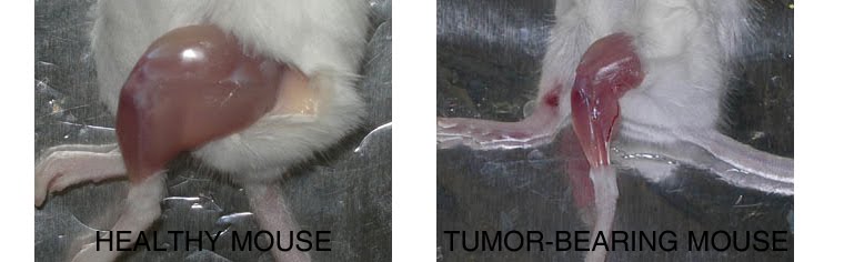
Compared to a control mouse (left) a tumor-bearing mouse (right) displays a dramatic muscle wasting. This loss of muscle mass is called cancer cachexia.
Exogenous gene expression in regenerating muscle
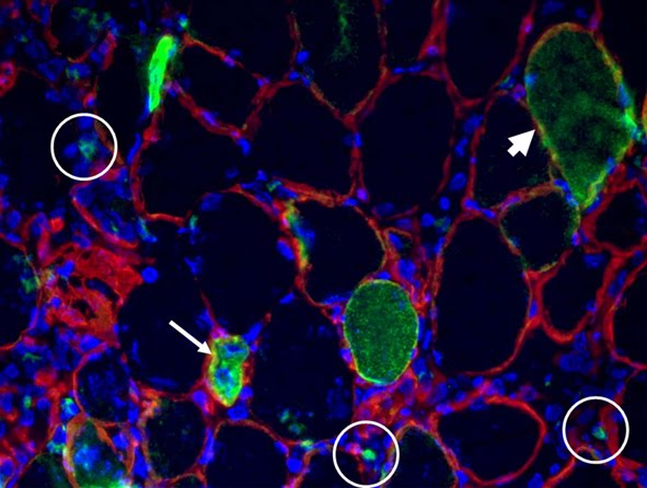
Depicted here is the over-expression of Green Fluorescent Protein (GFP, green; click on the image to access Tsien's Lab) in interstitial cells (circled), nascent myofibers (arrow) and adult fibers (arrowhead), in a regenerating Tibialis Anterior following focal injury. Laminin staining (red) highlights the basement membrane surrounding the skeletal muscle tissue, while nuclei are stained in blue. We do gene delivery by electroporation to study the regulation of muscle regeneration.
Cultures of myotubes on a conductive surface in a parallel orientation.
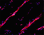
C2C12 cells cultured on gold, by mean of adhesion to 100 nm-wide stripes coated with anti Stem Cell antigen1 (Sca1) Ab. Nuclei (blue) and actin cytoskeleton (red) staining highlights the selective cells adhesion on the Ab-coated stripes and the formation of parallel multinucleated syncytia (myotubes).
8/31/2010
LAB METHODS: isolation of skeletal muscle fibers
The procedure after Dr. Peter Zammitt's...linked here are the figures of our version of the method
LAB METHODS: the simplest mycoplasma test

Mycoplasma is a common contaminant of cell cultures throughout the wolrd. It affects experimental results and it perturbs cell behaviour. Mycoplasma is a genus of bacteria that lack a cell wall.Without a cell wall, they are unaffected by many common antibiotics such as penicillin or other beta-lactam antibiotics that target cell wall synthesis. It is very difficult to get rid of mycoplasma: the best is to screen regularly for contamination and to throw out the contaminated cells! Here is the method for the screening....in italian
RELOCATING IN PARIS - UPMC

NEWS! Effective September the 1st I am at the University Paris VI Pierre et Marie Curie. I do teaching and research as Maître de Conférences. When I left Italy for the USA I had in my luggage a bottle of Barolo Granbussia 1982 by Aldo Conterno. It was a great experience - both the wine and postdoc! This time, coming to Paris, I put in my bag a Barolo Vigneto Arborina 2005 by Elio Altare - thanks Elio! I got to teach something to these guys...
7/30/2010
ARTICLES: link to Aulino et al. BMC Cancer 2010
 In the paper linked above we perform an extensive molecular, cellular and physiological characterization of the cancer cachexia-inducing C26 colon carcinoma in mouse. This widely diffesed murin model of cancer and cancer associated cachexia was originally developed by Corbett et al. in 1975, during an effort to establish an animal colon tumor model for biological and chemotherapy studies. Colon tumors were induced and transplanted in different inbred mouse strains. Four tumors survived the first transplant, which displayed a variety of histological and malignancy features. These four tumors included the colon tumor 26, described as an undifferentiated Grade IV carcinoma.
Since then, the model spread around the world and was exploited for a pletora of studies in the absence of an organic description of its main features.This gave rise to misunderstandings and mistakes, such as describing the C26 tumor as an adenocarcinoma without showing histological images.Today, the communities of scientists exploiting the C26 model to study either cancer or cachexia are not fully aware of each other’s works (as shown by cross-reference analysis on PubMed) and this may have deleterious consequences for the progress of integrative medicine applied to a complex syndrome associated with underlying illness. We suggest that “C26” be included among the keywords whenever work is conducted on this experimental model to provide adequate visibility.
Among the NOVEL DATA that we shown in this paper, I'd like to stress the functional analysis of cachectic muscles. We found that while the drop in muscle force is a hallmark of cachexia, it is simply due to muscle atrophy. In fact, no differences exist between normalized force (i.e. specific force) of control and cachectic animals. On the other hand, murine cachectic muscles are characterized by fatigue, which is in agreement with clinical observations. Left: C26 H&E staining. The measures shown below refer to the EDL muscle of ctr and C26-bearing mice.

6/30/2010
CLASSES, LECTURES ETC: Materials for the students of Tissue engineering School of Dentistry
Attached to the link there is a summary (in Italian) of basic concepts of Tissue engineering for the sutudents of the School of Dentistry.
IN allegato - link nel titolo - una dispensa con i concetti base di ingegneria tissutale presentati nell'ADE per gli studenti di OPD.
Linked to this text there are several articles on this topic.
A review...
IN allegato - link nel titolo - una dispensa con i concetti base di ingegneria tissutale presentati nell'ADE per gli studenti di OPD.
Linked to this text there are several articles on this topic.
A review...
6/24/2010
LAB METHODS: 3D acellular scaffold from skeletal muscle
The method linked here is a simplified procedure to obtain an acellular scaffold suitable for cell culture of myoblasts into a 3D scaffold.


5/26/2010
Cachexia, sarcopenia, inactivity: the three Fates (Moirae) of muscle atrophy
A common question is: what is the difference, if any, between muscle atrophy conditions which look all similar? What happens to our muscles when one lays in bed with a brokne leg, as opposed to getting cancer or happily ageing? Dr. Evans, one of the wolrd experts on muscle atrophy, simply and concisely pinpoints the differences in this review...

from:
Am J Clin Nutr. 2010 Apr;91(4):1123S-1127S. Epub 2010 Feb 17.
Skeletal muscle loss: cachexia, sarcopenia, and inactivity.
by Evans WJ.
Division of Geriatrics, Department of Medicine, Duke University Medical Center, Durham, NC 27709, USA. william.j.evans@gsk.com

from:
Am J Clin Nutr. 2010 Apr;91(4):1123S-1127S. Epub 2010 Feb 17.
Skeletal muscle loss: cachexia, sarcopenia, and inactivity.
by Evans WJ.
Division of Geriatrics, Department of Medicine, Duke University Medical Center, Durham, NC 27709, USA. william.j.evans@gsk.com
5/25/2010
LAB METHODS: CMFDA cell tracker

The protocol linked here is a method to label live cells and assess whether they are VITAL and/or STRESSED. The dye in fact accumulates inside the cells due to a chemical modification that makes it non-cell permeant. This esterase reaction requires glutathione. Therefore a shift toward low fluorescence characterizes a cell population with depleted levels of glutathione, i.e. that have been subjected to oxidative stress - GSH being the principal buffer of the redox state.
5/24/2010
caveat for the use of the Molecular Probes anti-Mouse Alexa Fluor 350 Ab
Linked here is the comparison between two Molecular Probes secondary antibodies: anti-M AF350-conjugated vs the classical AF488-conjugated. Thanks Paola for trying...
Looking at something in blue is cool but rather blues!
Looking at something in blue is cool but rather blues!
Il caso e la necessità, Le hasard et la nécessité, Chance and Necessity
"Tutto ciò che siste nell'universo è il frutto del caso e della necessità"
"Tout ce qui existe dans l'univers est le fruit du hasard et de la nècessité"
"Everything in the universe is the fruit of chance and necessity"
Democritus, circa 460-370 BC
IN: Jacques Monod, Le Hasard et al nécessité: Assai sur la phyolosophie naturelle.

Léon-Alexandre Delhomme (1841-1895)
Description
Democritus che medita sull'anima, bronzo, 1868.
Démocrite méditant sur le siège de l'âme, bronze, 1868.
Democritus meditating on the seat of the soul, bronze, 1868.
5/19/2010
CLASSES, LECTURES ETC: Regenerative Medicine & Tissue Engineering
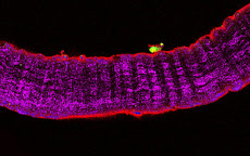
Here is the link to the ENGLISH VERSION of an introductory lecture to tissue engineering. The principles of the latter are summarized, and various strategies for regenerative medicine are presented through discussion of the most recent outbreaking reports in the field. For the 1st y medical students and biotechnology students. Talking about students, I have to acknowledge the great work done in the lab by Barbara Perniconi and Alessandra Costa (some of their data on acellular skeletal muscle are shown). A similar, broader lesson on regenerative medicine in FRENCH has been published here.

Figure legend: confocal image of a 20 micron tick cryosection of a murine acellular skeletal muscle matrix (laminin staining, red)

Figure legend: confocal image of a 20 micron tick cryosection of a murine acellular skeletal muscle matrix (laminin staining, red)
1/08/2010
A flu dealed with a secret treatment - article by Nerina Dirindin
Linked to the tile is an article, published by our great online magazine Lavoce.info, on the incredible management of the flu crisis by the Italian Minister of Health. The article is in Italian, but I wish to summarize it in English asap. Below is a little taste of the content...
About 4% of the Italian population has been vaccinated against the flu virus, which has recenlty given rise to pandemic emergency. On the other hand, the Italian government has bought from Novartis viruses for 40% of the population. This is not the real issue, though (except for singificant stocking and expiration problems). What is unbeliavable, is that the Ministry of Health has purchased the vaccines by using a procedure allowed by the Prime Minister (ordinanza n. 3275 del presidente del Consiglio del 2003) for cases of risk of terrorist attack. This procedure is secret, direct and charges almost all the burden on the government rather than on the company - the criticism comes from our Corte dei Conti (the top organ of suveillance of public expenses). For istance, the government will take care for all the expenses due to adverese effects or aother complications in the population underging vaccination. To date, it is not clear the cost of such operation.
About 4% of the Italian population has been vaccinated against the flu virus, which has recenlty given rise to pandemic emergency. On the other hand, the Italian government has bought from Novartis viruses for 40% of the population. This is not the real issue, though (except for singificant stocking and expiration problems). What is unbeliavable, is that the Ministry of Health has purchased the vaccines by using a procedure allowed by the Prime Minister (ordinanza n. 3275 del presidente del Consiglio del 2003) for cases of risk of terrorist attack. This procedure is secret, direct and charges almost all the burden on the government rather than on the company - the criticism comes from our Corte dei Conti (the top organ of suveillance of public expenses). For istance, the government will take care for all the expenses due to adverese effects or aother complications in the population underging vaccination. To date, it is not clear the cost of such operation.
Subscribe to:
Posts (Atom)
THE NETWORK OF OUR COLLABORATORS 2017

We collaborate with the Myology Group and the Cochin Hospital in Paris for stem cell studies and SRF, with the Cancer Centre at Ohio State University, Columbus for studies on the mechanisms underlying cachexia, with the Neurorehabilitation Unit at University of Pisa for clinical studies, with Pharmacology and Bioinformatics at the University of Urbino for advanced statistical analyses, with the Anatomy Section at the University of Perugia and with GYN/OB at the University of Western Piedmont for studies related to circulating factors and myogenic cell responses in cachexia, with the Biotech-Med Unit at ENEA, Chemistry in Rome and Anatomy in palermo for tissue engineering applications. Functional studies are carried out in our Departement in Rome in collaboration with Musaro's laboratory.


