RESEARCH INTERESTS: Cellular and molecular mechanisms of striated muscle physiopathology

1. PHARMACOLOGICAL, PHYSICAL, AND NUTRITIONAL INTERVENTIONS AGANIST CANCER CACHEXIA: My laboratory is focused on different approaches to counteract cancer cachexia, including pharmacological (exercise mimetics), physiological (physical activity), and nutritional (supplements) interventions in humans and animal models. 2. MYOFIBER MEMBRANE DAMAGE AND REPAIR: Duchenne Muscular Dystrophy (DMD), is a lethal genetic, muscle-wasting disease, characterized by progressive muscle fragility and weakness. The muscle membrane repair mechanism (MRM) is an active resealing pathway involving vesicle-sarcolem fusion to “patch” the compromised plasma membrane and represents a possible target to counteract muscle wasting in DMD, in which the chronic cycle of muscle degeneration-regeneration plays a pivotal role in disease progression. 3. PATENTS AND TECHNOLOGY TRASNFER: I am co-inventor of a patented procedure to produce Hsp60-enriched exosomes with exercise-mimetic activity, a product that is, therefore, called Physiactisome. Patent: Physiactisome – «Procedure for the synthesis of HSP-containing exosomes and their use against muscle atrophy and cachexia» - patent n. 102018000009235 on 8/10/2018, deposited by Università di Palermo. Owners: Università di Palermo, Università di Roma La Sapienza, Nanovector Torino, Sorbonne Université. List of inventors: Valentina Di Felice, Rosario Barone, Antonella Marino Gammazza, Campanella Claudia, Cappello Francesco, Farina Felicia, Eleonora Trovato, Daniela D’Amico, Filippo Macaluso, Dario Coletti, Sergio Adamo, Gabriele Multhoff, Paolo Gasco. International publication number WO 2020/075004 A1. This product can be exploited against muscle atrophy, since it ameliorates muscle endurance and homeostasis. The presentation of the product and the corresponding Spinoff project (iBioTHEx) was awarded the third prize at the EIT JumpStarter Grand final, Riga, Latvia, 15-17/11/2019, Health category. 4. PHYSIOPATHOLOGY OF MUSCLE TISSUES: I contribute to discovering and explaining those mechanisms underlying pathologies of the striated and smooth muscle tissues; this activity is carried out at Sorbonne University by using genetic murine models.

Cancer cachexia

Cancer cachexia
Compared to a control mouse (left) a tumor-bearing mouse (right) displays a dramatic muscle wasting. This loss of muscle mass is called cancer cachexia.

Exogenous gene expression in regenerating muscle

Exogenous gene expression in regenerating muscle
Depicted here is the over-expression of Green Fluorescent Protein (GFP, green; click on the image to access Tsien's Lab) in interstitial cells (circled), nascent myofibers (arrow) and adult fibers (arrowhead), in a regenerating Tibialis Anterior following focal injury. Laminin staining (red) highlights the basement membrane surrounding the skeletal muscle tissue, while nuclei are stained in blue. We do gene delivery by electroporation to study the regulation of muscle regeneration.

RESEARCH INTERESTS: Tissue engineering of skeletal muscle

Background and rationale.

Tissue engineering lies at the interface of regenerative medicine and developmental biology, and represent an innovative and multidisciplinary approach to build organs and tissues (Ingber and Levin, Development 2007). The skeletal muscle is a contractile tissue characterized by highly oriented bundles of giant syncytial cells (myofibers) and by mechanical resistance. Contractile, tissue-engineered skeletal muscle would be of significant benefit to patients with muscle deficits secondary to congenital anomalies, trauma, or surgery. Obvious limitations to this approach are the complexity of the musculature, composed of multiple tissues intimately intermingled and functionally interconnected, and the big dimensions of the majority of the muscles, which imply the involvement of an enormous amount of cells and rises problems of cell growth and survival (nutrition and oxygen delivery etc.). Two major approaches are followed to address these issues. Self-assembled skeletal muscle constructs are produced in vitro by delaminating sheets of cocultured myoblasts and fibroblasts, which results in contractile cylindrical “myooids.” Matrix-based approaches include placing cells into compacted lattices, seeding cells onto degradable polyglycolic acid sponges, seeding cells onto acellularized whole muscles, seeding cells into hydrogels, and seeding nonbiodegradable fiber sheets. Recently, decellularized matrix from cadaveric organs has been proven to be a good scaffold for cell repopulation to generate functional hearts in mice (Ott et al. Nature Medicine 2008).

I have obtained cultures of skeletal muscle cells on conductive surfaces, which is required to develop electronic device–muscle junctions for tissue engineering and medical applications1. I aim to exploit this system for either recording or stimulation of muscle cell biological activities, by exploiting the field effect transistor and capacitor potential of the conductive substratum-cell interface. Also, we are able to create patterned dispositions of molecules and cells on gold, which is important to mimic the highly oriented pattern myofibers show in vivo.

I have found that Static magnetic fields enhance skeletal muscle differentiation in vitro by improving myoblast alignment2. Static magnetic field (SMF) interacts with mammal skeletal muscle; however, SMF effects on skeletal muscle cells are poorly investigated. 80 +/- mT SMF generated by a custom-made magnet promotes myogenic cell differentiation and hypertrophy in vitro. Finally, we have transplanted acellular scaffolds to study the in vivo response to this biomaterial3, which we want to exploit for tissue culture and regenerative medicine of skeletal muscle.

The specific aims of my current research are:

1) to increase and optimize the production and alignment of myogenic cells and myotubes in vitro;

2) to manipulate the niche of muscle stem cells aimed at ameliorating their regenerative capacity in vivo;

3) to develop muscle-electrical devices interactions. We plan to exploit the cell culture system on conductive substrates for either recording or stimulation of muscle cell biological activities, by exploiting the field effect transistor and capacitor potential of the conductive substratum-cell interface.

4) to better clarify the biological effects of Static Magnetic Fields. With the aim to characterize the molecular mechanism underlying the effects of SMF on cell differentiation and alignment we are exposing molecules and cells to SMF below 1T.
5)
to produce pre-assembled, off-the-shelf skeletal muscle. We are seeding acellularized muscle scaffold with various cell types, with the goal to obtain functional muscle with vascular supply and nerves.


REFERENCES

1) Coletti D. et al., J Biomed Mat Res 2009; 91(2):370-377.


2) Coletti D. et al., Cytometry A. 2007;71(10):846-56.


3) Perniconi B. et al. Biomaterials, 2011 in press

Cultures of myotubes on a conductive surface in a parallel orientation.

Cultures of myotubes on a conductive surface in a parallel orientation.
C2C12 cells cultured on gold, by mean of adhesion to 100 nm-wide stripes coated with anti Stem Cell antigen1 (Sca1) Ab. Nuclei (blue) and actin cytoskeleton (red) staining highlights the selective cells adhesion on the Ab-coated stripes and the formation of parallel multinucleated syncytia (myotubes).

12/15/2009

Methodological papers: my favourite hits


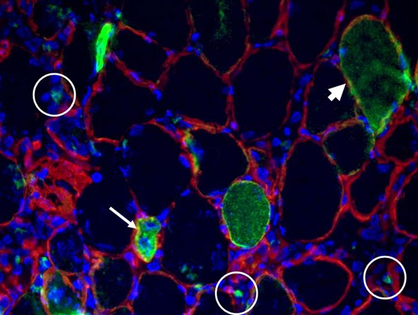
FIGURE LEGEND: Cross-sectional view of a Tibialis Anterior injected with GFP DNA (green), electroporated in our Roman lab following Donà et al., and immunostained for laminin (red).


Some times a scientific article is outstanding because of its breakthrough findings, some times it is particularly elegant in its demonstrations, some other times an article is just so useful! The following are some of my favourite methodological papers.

For people working on gene delivery by electroporation to skeletal muscle tissue, Donà et al. carefully address which are the best settings for the square wave electroporator. The efficiency of this approach can be pretty high, as shown by this article and by the image above. We often perform electroporation-mediated delivery of two different plasmids with the aim to obtain co-expression. Which is the correct ratio between the two constructs? For instance, if we want to co-express Green Fluorescent Protein (GFP) as an expression marker, which amount of the plasmid of interest must we combine with the GFP to be sure that it is expressed by the fibers that turn out green? Rana et al. have found that co-expression rate between BFP and GFP in skeletal muscle is about 100% in their experimental settings.

A caveat on the use of GFP comes from this paper by Goodell and co-workers: they explain why skeletal muscle fiber-specific green autofluorescence can generate potential artifacts and show how to discriminate between autofluorescence and GFP signal. There is a (fake) green side and a dark side of GFP, i.e. the fact that its expression interferes with polyubiquitination. Those working on proteasome-mediated protein degradation should be very careful when ectopically expressing GFP, as shown by Baens et al.

To assess cell damage it is possible to exploit Evans Blue Dye, which is cell-impermeant unless the plasma membrane is damaged. A complete characterization of its use to label muscle fiber damage is described by Hamer et al. They tell you everything you wanted to know on EBD and you never dared to ask.

YACs, BACs, PACSs…are they Dr. Seuss’ creatures or carriers for successful generation of transgenic mice when transfer of large fragments of cloned genomic DNA is needed? Giraldo and Motoliu will tell you.

When we deal with myogenic cell cultures, we often obtain a mixed population of myotubes that have differentiated in the presence of a residual population of unfused myoblasts, i.e. syncitia vs single cells. The paper by Kitzmann et al. is not only a great JCB paper on cell cycle-dependent regulation of MyoD and Myf5 but also provides a trick to separate myotubes from myoblasts.

It makes no sense that we produce data if we do not know how to handle or to communicate them. I was struck by the news that about 50% of scientific articles contain errors of data analysis or reporting. C.H. Olsen reviews the use of statistics in articles published – yes, published – in Infection and Immunity and discusses the most common mistakes.

No comments:

Post a Comment

THE NETWORK OF OUR COLLABORATORS 2017

THE NETWORK OF OUR COLLABORATORS 2017
We collaborate with the Myology Group and the Cochin Hospital in Paris for stem cell studies and SRF, with the Cancer Centre at Ohio State University, Columbus for studies on the mechanisms underlying cachexia, with the Neurorehabilitation Unit at University of Pisa for clinical studies, with Pharmacology and Bioinformatics at the University of Urbino for advanced statistical analyses, with the Anatomy Section at the University of Perugia and with GYN/OB at the University of Western Piedmont for studies related to circulating factors and myogenic cell responses in cachexia, with the Biotech-Med Unit at ENEA, Chemistry in Rome and Anatomy in palermo for tissue engineering applications. Functional studies are carried out in our Departement in Rome in collaboration with Musaro's laboratory.