RESEARCH INTERESTS: Cellular and molecular mechanisms of striated muscle physiopathology

1. PHARMACOLOGICAL, PHYSICAL, AND NUTRITIONAL INTERVENTIONS AGANIST CANCER CACHEXIA: My laboratory is focused on different approaches to counteract cancer cachexia, including pharmacological (exercise mimetics), physiological (physical activity), and nutritional (supplements) interventions in humans and animal models. 2. MYOFIBER MEMBRANE DAMAGE AND REPAIR: Duchenne Muscular Dystrophy (DMD), is a lethal genetic, muscle-wasting disease, characterized by progressive muscle fragility and weakness. The muscle membrane repair mechanism (MRM) is an active resealing pathway involving vesicle-sarcolem fusion to “patch” the compromised plasma membrane and represents a possible target to counteract muscle wasting in DMD, in which the chronic cycle of muscle degeneration-regeneration plays a pivotal role in disease progression. 3. PATENTS AND TECHNOLOGY TRASNFER: I am co-inventor of a patented procedure to produce Hsp60-enriched exosomes with exercise-mimetic activity, a product that is, therefore, called Physiactisome. Patent: Physiactisome – «Procedure for the synthesis of HSP-containing exosomes and their use against muscle atrophy and cachexia» - patent n. 102018000009235 on 8/10/2018, deposited by Università di Palermo. Owners: Università di Palermo, Università di Roma La Sapienza, Nanovector Torino, Sorbonne Université. List of inventors: Valentina Di Felice, Rosario Barone, Antonella Marino Gammazza, Campanella Claudia, Cappello Francesco, Farina Felicia, Eleonora Trovato, Daniela D’Amico, Filippo Macaluso, Dario Coletti, Sergio Adamo, Gabriele Multhoff, Paolo Gasco. International publication number WO 2020/075004 A1. This product can be exploited against muscle atrophy, since it ameliorates muscle endurance and homeostasis. The presentation of the product and the corresponding Spinoff project (iBioTHEx) was awarded the third prize at the EIT JumpStarter Grand final, Riga, Latvia, 15-17/11/2019, Health category. 4. PHYSIOPATHOLOGY OF MUSCLE TISSUES: I contribute to discovering and explaining those mechanisms underlying pathologies of the striated and smooth muscle tissues; this activity is carried out at Sorbonne University by using genetic murine models.

Cancer cachexia

Cancer cachexia
Compared to a control mouse (left) a tumor-bearing mouse (right) displays a dramatic muscle wasting. This loss of muscle mass is called cancer cachexia.

Exogenous gene expression in regenerating muscle
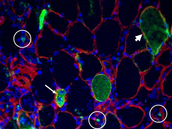
Exogenous gene expression in regenerating muscle
Depicted here is the over-expression of Green Fluorescent Protein (GFP, green; click on the image to access Tsien's Lab) in interstitial cells (circled), nascent myofibers (arrow) and adult fibers (arrowhead), in a regenerating Tibialis Anterior following focal injury. Laminin staining (red) highlights the basement membrane surrounding the skeletal muscle tissue, while nuclei are stained in blue. We do gene delivery by electroporation to study the regulation of muscle regeneration.

RESEARCH INTERESTS: Tissue engineering of skeletal muscle

Background and rationale.

Tissue engineering lies at the interface of regenerative medicine and developmental biology, and represent an innovative and multidisciplinary approach to build organs and tissues (Ingber and Levin, Development 2007). The skeletal muscle is a contractile tissue characterized by highly oriented bundles of giant syncytial cells (myofibers) and by mechanical resistance. Contractile, tissue-engineered skeletal muscle would be of significant benefit to patients with muscle deficits secondary to congenital anomalies, trauma, or surgery. Obvious limitations to this approach are the complexity of the musculature, composed of multiple tissues intimately intermingled and functionally interconnected, and the big dimensions of the majority of the muscles, which imply the involvement of an enormous amount of cells and rises problems of cell growth and survival (nutrition and oxygen delivery etc.). Two major approaches are followed to address these issues. Self-assembled skeletal muscle constructs are produced in vitro by delaminating sheets of cocultured myoblasts and fibroblasts, which results in contractile cylindrical “myooids.” Matrix-based approaches include placing cells into compacted lattices, seeding cells onto degradable polyglycolic acid sponges, seeding cells onto acellularized whole muscles, seeding cells into hydrogels, and seeding nonbiodegradable fiber sheets. Recently, decellularized matrix from cadaveric organs has been proven to be a good scaffold for cell repopulation to generate functional hearts in mice (Ott et al. Nature Medicine 2008).

I have obtained cultures of skeletal muscle cells on conductive surfaces, which is required to develop electronic device–muscle junctions for tissue engineering and medical applications1. I aim to exploit this system for either recording or stimulation of muscle cell biological activities, by exploiting the field effect transistor and capacitor potential of the conductive substratum-cell interface. Also, we are able to create patterned dispositions of molecules and cells on gold, which is important to mimic the highly oriented pattern myofibers show in vivo.

I have found that Static magnetic fields enhance skeletal muscle differentiation in vitro by improving myoblast alignment2. Static magnetic field (SMF) interacts with mammal skeletal muscle; however, SMF effects on skeletal muscle cells are poorly investigated. 80 +/- mT SMF generated by a custom-made magnet promotes myogenic cell differentiation and hypertrophy in vitro. Finally, we have transplanted acellular scaffolds to study the in vivo response to this biomaterial3, which we want to exploit for tissue culture and regenerative medicine of skeletal muscle.

The specific aims of my current research are:

1) to increase and optimize the production and alignment of myogenic cells and myotubes in vitro;

2) to manipulate the niche of muscle stem cells aimed at ameliorating their regenerative capacity in vivo;

3) to develop muscle-electrical devices interactions. We plan to exploit the cell culture system on conductive substrates for either recording or stimulation of muscle cell biological activities, by exploiting the field effect transistor and capacitor potential of the conductive substratum-cell interface.

4) to better clarify the biological effects of Static Magnetic Fields. With the aim to characterize the molecular mechanism underlying the effects of SMF on cell differentiation and alignment we are exposing molecules and cells to SMF below 1T.
5)
to produce pre-assembled, off-the-shelf skeletal muscle. We are seeding acellularized muscle scaffold with various cell types, with the goal to obtain functional muscle with vascular supply and nerves.


REFERENCES

1) Coletti D. et al., J Biomed Mat Res 2009; 91(2):370-377.


2) Coletti D. et al., Cytometry A. 2007;71(10):846-56.


3) Perniconi B. et al. Biomaterials, 2011 in press

Cultures of myotubes on a conductive surface in a parallel orientation.
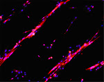
Cultures of myotubes on a conductive surface in a parallel orientation.
C2C12 cells cultured on gold, by mean of adhesion to 100 nm-wide stripes coated with anti Stem Cell antigen1 (Sca1) Ab. Nuclei (blue) and actin cytoskeleton (red) staining highlights the selective cells adhesion on the Ab-coated stripes and the formation of parallel multinucleated syncytia (myotubes).

12/20/2015

Miro painting by Transmission Electron Microscopy


Electrophotomicrograph of murine skeletal muscle extra-cellular matrix. Collagen fibers and debris, likely deriving from cell damage or necrosis are visible.

12/16/2015

Demonstration that feijoada fits better with beer than Bordeaux wine


Introduction Feijoada is a stew of beans with beef and pork,which is the national Brazilian dish. It is served with rice and farofa (manioc flour sauted in butter). "Brazilian feijoada (feijoada brasileira) is prepared with black beans,[3] a variety of salted pork or beef products, such as pork[3] trimmings (ears, tail, feet),[3] bacon, smoked pork ribs, and at least two types of smoked sausage and jerked beef (loin and tongue) and in some regions of the northeast, like Bahia and Sergipe they usually add vegetables like cabbage, kale, potatoes, carrots, okra, pumpkin, chayote and sometimes banana. They are added at the end of the cooking, on top of the meat, so they are cooked by the vapors of the beans and meat stew. This stew is best prepared over low fire in a thick clay pot. The final dish has the beans and meat pieces barely covered by a dark purplish-brown broth. The taste is strong, moderately salty but not spicy, dominated by the flavors of black bean and meat stew. It is customary to serve it with white rice and oranges, the latter to help with digestion. Feijão com arroz is the rice and black beans without the addition of the meat." From wikipedia (1) Matherials and methods "Ingredients 2 tablespoons olive oil 2 cups chopped onions 2 tablespoons chopped garlic 2 bay leaves Salt Freshly ground black pepper 1 pound choriço sausage, sliced 1/4-inch thick 1 pound carne seca or other salted cured beef, soaked overnight and cubed 1 pound baby back spareribs, cut into individual ribs 1 pound black beans 10 cups water 4 cups collared or kale greens, sauteed in olive oil 4 cups cooked white rice Brazilian hot sauce Garnish: 1 orange, halved and cut into thin slices, and Farofa Directions In a large heavy-bottom saucepan, over medium heat, add the oil. When the oil is hot, add the onions and garlic. Crush the bay leaves and add to the pan. Season with salt and pepper. Saute for 5 minutes. Add the sausage. Continue to cook for 4 minutes. Add the cubed beef, ribs, beans and water. Bring the liquid to a boil, reduce the heat to medium low and simmer until the beans are tender, about 2 1/2 hours. Adding water as necessary to keep the beans covered. Using the back of a ladle, mash 1/4 of the beans. Reseason with salt and pepper if needed. To serve, spoon some of the greens and rice onto each serving plate. Spoon the Feijoada over the rice. Shake some of the hot sauce over the entire plate. Garnish with the orange slices and farofa." Cited from SmithsonianMag (2). Results We tasted home made fejioada pairing with either lager beer or red bordeaux wine. The panel was composed by about twelve lab members, specifically trained to wine and food tasting by dozens of lab parties, with an experience of several years. 85% of the above population agreed that beer matches better than red wine to the dish. In particular, we noted that beer CO2 is sufficient to equilibrate the fatty sauce while respecting the light body of the rise and manioc side dishes. Discussion Here we compared the traditional pairing of fejioada with lager beer to a more innovative approach based on an equilibrate blend represented by the French bordeaux wine. We concluded that beer is more appropriate than red wine to accompany the dish. Since light beer was slightly inadequate to support the full bodied and very aromatic meat sauce, we foresee that champagne wine might have the right combination of aromatic intensity and persistence and body, not to forget effervescence, to perfectly pair fejioada. This hypothesis grunts further investigation. Figure
Acknoledgements Emidio Matos is gratefully acknoledged for the fejioada and farofa preparation. References 1) https://en.wikipedia.org/wiki/Feijoada 2) http://www.smithsonianmag.com/arts-culture/Celebrate-Brazil-with-Emerils-Feijoada-180951699/?no-ist

Christmas 2015 in the lab


12/01/2015

METHODS: cell culture conditions for C2C12, L6 cells, C26 and LLC etc.


We have a few cell lines available in the lab, including the following widely diffused cells: fibroblasts, C2C12 murine myoblasts, L6 rat myoblasts, H9C2 cardiomyocytes, C26 murine colon carcinoma, Lewis Lung murine carcinoma. Linked here are some notes and information about line maintenance and myogenic differentiaiton.

9/25/2015

Focus: Biomaterials and bioactive molecules to drive differentiation in striated muscle tissue

Below the point on our most recent research in Tissue enginnering. Recently, an article by Armstrong et al. entitled « TOP 10 DEVELOPMENTS IN STEM CELL BIOLOGY OVER THE LAST 30 YEARS » summarized ten quantum leaps in stem cells research. These are: 1) THE DISCOVERY AND ISOLATION ADULT STEM CELLS 2) THE FIRST EMBRYONIC STEM CELLS FROM MICE 3) THE DEVELOPMENT OF MAMMALIAN CLONING 4) HUMAN EMBRYONIC STEM CELLS 5) THE CONCEPT OF CANCER STEM CELLS 6) INDUCED PLURIPOTENT STEM CELLS 7) MESENCHYMAL STEM CELLS 8) THE TISSUE ENGINEERING WITH STEM CELLS 9) THE IMPROVEMENT OF GENETIC MANIPULATION THE BEGINNING OF TRANSLATIONAL MEDICAL APPLICATIONS (In: Armstrong L. et al. STEM CELLS 2012;30:2–9. link fo tull text: https://drive.google.com/file/d/0B_z0HQaQV25yX3EzOTVBcXNSVTA/view?usp=sharing) We have cited in this article (Perniconi et al. Biomaterials, 2011) as an outstanding example of tissue engineering approach. That's great! Thank you. We mainly focus on the use of Biomaterials to drive differentiation in striated muscle tissue. The latter has a peculiar way of regeneraitng (Restoration versus reconstruction: cellular mechanisms of skin, nerve and muscle regeneration compared. Coletti D, Teodori L, Lin Z, Beranudin JF, Adamo S. Regen Med Res. 2013 Oct 1;1(1):4. doi: 10.1186/2050-490X-1-4. eCollection 2013 Dec. Review. PMID: 25984323. Full text here). Advances in tissue replacement and regeneraiton involve muscle replacement, such as in the case of volumetric muscle loss, and inflammation control, such as in myopathies characterized by chronic inflammation (Inflammation in muscle repair, aging, and myopathies. Bouché M, Muñoz-Cánoves P, Rossi F, Coletti D. Biomed Res Int. 2014;2014:821950. doi: 10.1155/2014/821950. Epub 2014 Aug 4. No abstract available. PMID: 25162030. Full text here). Of the various alternative approaches to replace or regenerate muscle, the use of biomaterials has gained major attention (Biomaterials and bioactive molecules to drive differentiation in striated muscle tissue engineering. Di Felice V, Forte G, Coletti D. Front Physiol. 2015 Feb 23;6:52. doi: 10.3389/fphys.2015.00052. eCollection 2015. PMID: 25755644. Full text here). In particular, scaffolds obtained by decellularization of small intestinal submucosa(SIS),urinary bladder mucosa(UB) and skeletal muscle are getting very promising results at pre-clinical and clinical level (reviewed in Native extracellular matrix: a new scaffolding platform for repair of damaged muscle. Teodori L, Costa A, Marzio R, Perniconi B, Coletti D, Adamo S, Gupta B, Tarnok A. Front Physiol. 2014 Jun 16;5:218. doi: 10.3389/fphys.2014.00218. eCollection 2014. Review. PMID: 24982637. Full text here). A consistent body of evidence indicates that extra-cellular matrix (ECM) proteins regulate stem cell differentiation and renewal and are highly relevant to tissue engineering applications. The ECM also provides a supportive medium for blood or lymphatic vessels and for nerves. Thus, the ECM is the nature's ideal biological scaffold material. We have been focusing on ECM derived from decellularized skeletal muscle. This muscle acellular scaffold (MAS) may represent a suitable environment for muscle and non-muscle 3D constructs characterized by a highly organized structure whose relative stability promotes integration with the surrounding tissues. (Muscle acellular scaffold as a biomaterial: effects on C2C12 cell differentiation and interaction with the murine host environment. Perniconi B, Coletti D, Aulino P, Costa A, Aprile P, Santacroce L, Chiaravalloti E, Coquelin L, Chevallier N, Teodori L, Adamo S, Marrelli M, Tatullo M. Front Physiol. 2014 Sep 26;5:354. doi: 10.3389/fphys.2014.00354. eCollection 2014. PMID: 25309452. Full text here). Our recent work also highlights the plasticity of MAS, suggesting that it may be possible to consider MAS for a wider range of tissue engineering applications than the mere replacement of volumetric muscle loss. (Muscle extracellular matrix scaffold is a multipotent environment. Aulino P, Costa A, Chiaravalloti E, Perniconi B, Adamo S, Coletti D, Marrelli M, Tatullo M, Teodori L. Int J Med Sci. 2015 Apr 6;12(4):336-40. doi: 10.7150/ijms.10761. eCollection 2015. PMID: 25897295. Full text here). Nonetheless, caution is imposed when announcing these major progresses on skeletal muscle tissue engineering, since is still impossible to fully reconstruct such a highly hierarchized, big, and complex organ for in vivo transplantaiton. While for these ambitious in vivo tissue engineering applications, there may still be a long way to go, novel in vitro applications for tissue engineered contructs are emerging, such as 3D cultures aimed at better mimicking an in vivo models. It is self-evident that bidimensional cultures are very limited insofar as the physiological 3D tissue organization they yield is somewhat approximate. With the current need to develop experimentals models replacing or refining animal-based research, these ideas are becoming increasingly appealing. In conclusion, we believe that the best bet for skeletal muscle TE is to focus on specific, anatomically defined solutions or on 3D in vitro modeling of muscle tissue for basic and applied research (Skeletal muscle tissue engineering: best bet or black beast? Perniconi B, Coletti D. Front Physiol. 2014 Jul 4;5:255. doi: 10.3389/fphys.2014.00255. eCollection 2014. No abstract available. PMID: 25071600. Full text here). We are confident that we will eventually be able to transform the black beast (i.e., striated muscle tissue engineering) into the best bet (i.e., a successful clinical practice based on engineered muscles).

Figure from: Coletti Det al. Regen Med Res. 2013 Oct 1;1(1):4. Hematoxilin- and eosin-staining (H&E) and immunofluorescence localization of muscle fiber damage (red) and of the membrane basement component laminin (green) on serial cross-sections of murine Tibialis anterior muscle (only a portion of the muscle is shown). Thirty minutes before fixation, the muscle was subjected to two types of physical injury: mechanical stress by crunching and tearing with forceps (LEFT) and freezing by applying a liquid nitrogen-cooled steel forceps to the surface (facing down in the picture) for 10 seconds (CENTER). Apart for the edema and fiber swelling visible in the images on the right, no major alterations of the basement membrane are seen following focal injury. In mice injected with Evans Blue Dye (EBD, RIGHT), injury muscle fiber necrosis (red) is apparent 8 h after freezing thanks to accumulation of EBD in the interior part of the damaged fibers. The muscle fibers die and are either renewed or replaced within the intact scaffold represented by the membrane basement, which wraps each fiber.Note that in all cases the basement membrane remains intact in sharp contrast with muscle fiber damage, therefore the general architecture of the muscle is preserved.

THE NETWORK OF OUR COLLABORATORS 2017

THE NETWORK OF OUR COLLABORATORS 2017
We collaborate with the Myology Group and the Cochin Hospital in Paris for stem cell studies and SRF, with the Cancer Centre at Ohio State University, Columbus for studies on the mechanisms underlying cachexia, with the Neurorehabilitation Unit at University of Pisa for clinical studies, with Pharmacology and Bioinformatics at the University of Urbino for advanced statistical analyses, with the Anatomy Section at the University of Perugia and with GYN/OB at the University of Western Piedmont for studies related to circulating factors and myogenic cell responses in cachexia, with the Biotech-Med Unit at ENEA, Chemistry in Rome and Anatomy in palermo for tissue engineering applications. Functional studies are carried out in our Departement in Rome in collaboration with Musaro's laboratory.